


| Journal of Clinical Gynecology and Obstetrics, ISSN 1927-1271 print, 1927-128X online, Open Access |
| Article copyright, the authors; Journal compilation copyright, J Clin Gynecol Obstet and Elmer Press Inc |
| Journal website http://www.jcgo.org |
Case Report
Volume 4, Number 3, September 2015, pages 279-281
Vulvar Granular Cell Tumor: A Rare Tumor in an Unusual Location
Isabel Barros Pereiraa, c, Ashfaq Khanb
aClinica Universitaria de Obstetricia e Ginecologia, CHLN - Hospital Universitario de Santa Maria, Faculdade de Medicina da Universidade de Lisboa, CAM - Centro Academico de Medicina de Lisboa, Portugal
bThe Whittington Hospital NHS Trust, Colposcopy and Vulva Disease Unit, London, UK
cCorresponding Author: Isabel Barros Pereira, Rua Joaquim Rocha Cabral, n.24, 4D 1600-086 Lisbon, Portugal
Manuscript accepted for publication August 25, 2015
Short title: Vulvar Granular Cell Tumor
doi: http://dx.doi.org/10.14740/jcgo350w
| Abstract | ▴Top |
Granular cell tumors are rare neoplasms of neural sheath origin, which rarely involve the vulvar area. We report the case of a 53-year-old postmenopausal woman who presented with an increasing vulvar mass with 2 years duration without associated symptoms. The physical examination revealed a hard, mobile nodular subdermal mass. An excision biopsy of the lesion was performed. The histopathologic exam revealed a granular cell tumor without features of malignancy. On the follow-up, 2 months after the excision, the patient was asymptomatic and presented a complete healed scar with no induration. Although granular cell tumors of the vulva are uncommon and mostly benign, they have a tendency for local recurrence. Once diagnosed with a granular cell tumor, the patient must be counseled to follow-up regularly.
Keywords: Vulvar tumor; Granular cell tumor; Follow-up
| Introduction | ▴Top |
Granular cell tumors (GCTs) are rare neoplasms of neural sheath origin, the majority of which occur in the skin, submucosal or subcutaneous tissue of head and neck, especially in the tongue and oral cavity (45-65%) [1, 2]. GCTs occur more often in females than males and in blacks than whites. The peak age incidence is in the fourth through fifth decades. In 98% of cases, these tumors are benign. The malignant form of the tumor is rare, highly aggressive and unresponsive to treatment [3, 4].
Vulvar involvement is rare and has been reported in 10% of cases. There have also been reported cases involving the ovary, uterus, cervix, vagina, mons pubis and episiotomy scar [1].
| Case Report | ▴Top |
We report the case of a 53-year-old postmenopausal woman who presented with an increasing vulvar mass with 2 years duration without associated symptoms. She presented no history of tenderness, discharge or any bleeding from the area. She denied history of previous vulvar lesion or systemic symptoms.
The physical examination revealed a hard, mobile nodular subdermal mass measuring 25 × 10 mm at the right labium major (Fig. 1). It was fixed to the overlying skin which had a normal appearance. The lesion was non-tender and on palpation there was no sign of discharge or bleeding. There were no palpable lymph nodes.
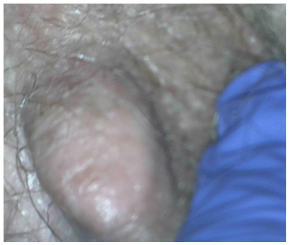 Click for large image | Figure 1. Hard, mobile nodular subdermal mass: clinical presentation of the vulvar granular cell tumor. |
An excision biopsy of the lesion was performed under general anesthesia. On macroscopic exam, the specimen measured 35 × 18 mm and had a surface raised skin covered area. The skin itself was unremarkable. On slicing, the cut surface was pale yellow and firm with some brown discoloration towards the deep edge focally. The changes extended to near or at the deep edge. The histopathologic exam revealed a circumscribed dermal tumor consisting of sheets of large, bland cells with voluminous granular cytoplasm. The cells had round and ovoid nuclei with prominent nucleoli. Immunohistochemistry showed that the tumor cells were S-100 positive and weakly CD68 positive. These features were those of a GCT without features of malignancy. The tumor was present at the deep excision margin.
On the follow-up appointment 2 months after the excision procedure, the patient was asymptomatic and presented a complete healed scar with no induration.
| Discussion | ▴Top |
GCTs are derived from neural tissue and immunohistochemical and ultrastructural evidence supports the current opinion that the tumor has its origin in Schwann cells [3]. Most cases are sporadic and the possibility of familial link needs further investigation [1].
On clinical presentation, GCTs are generally small (rarely larger than 4 cm in diameter), firm, solitary nodules lacking encapsulation and located in the subcutaneous layer. They are typically slow-growing and asymptomatic and can sometimes be confused with sebaceous cysts. Pruritus and pain have occasionally been reported. The mass is usually mobile and the overlying skin may be depigmented, occasionally ulcerated or even thickened [2]. Larger lesions may sometimes show an ulcerated surface, which may clinically give impression of a malignant neoplasm. The most common location of GCT in the female genital tract is the labium major.
There have been reports of GCT of the vulva, which are aggressive with multicentric or metastatic disease. The incidence of multicentric lesions ranges from 3% to 20% and this raises the suspicion of malignancy and possible association with regional or distant metastases [1, 2].
On cross-section, the tumor is poorly circumscribed with irregular margins and yellow-gray appearance.
Microscopically, GCTs are composed of loosely infiltrating sheets or clusters of large spindled cells. The main morphologic feature is the granularity of the cytoplasm caused by massive phagolysosomes accumulation. Nuclei are uniform, small and dark staining. In about half the cases, the squamous epithelium overlying the tumor shows pseudoepitheliomatous hyperplasia which may be mistaken for squamous carcinoma. Immunohistochemical stains are positive for S-100 protein, CD68, periodic acid Schiff, neuron specific enolase, peripheral nerve myelin proteins, vimentin and are diastase resistant.
Nuclear enlargement, hyperchromatism, pleomorphism, mitotic activity or increased cellularity are elements of the malignant variant of this tumor [4]. Malignant GCTs are often immunohistochemically negative for S-100 protein, neuron specific enolase and vimentin [4]. However, the distinction between benign and malignant GCT is difficult because of histologic similarity and lack of reliable criteria that can predict clinical behavior.
A third type of GCT has been described which has benign pathologic characteristics but behaves in a clinically malignant manner [4].
In our case, the tumor cells were S-100 positive and weakly CD68 positive, which suggests benignity.
The malignant form of the tumor may not be diagnosed until regional or distant metastasis occurs. Clinically, features associated with poor prognosis include rapid tumor growth, older age, tumor size more than 4 cm, vascular invasion, necrosis and local recurrence. This malignant variety is very aggressive with regional and metastatic spread and poor response to radiotherapy and chemotherapy [3]. Metastases can occur via lymphatic spread to regional lymph nodes and hematogeneous spread to liver, lungs and bones [3].
The differential diagnosis for GCT includes fibroma, lipoma, papilloma, hidradenoma, epidermal cyst, Bartholin cyst and melanoma. Unlike Schwannoma, there is no association with neurofibromatosis [3]. Schwannomas are also S-100 protein positive but are histologically completely different composed of spindle non-granular cells with compact and loose areas [3]. Immunochemical stains will help on the differential diagnosis [3].
The treatment of choice for all types is wide, local surgical excision [4]. In the malignant form of the tumor radical local surgery with a view for regional lymph node dissection should be carried out, if there are no distant metastases. Because the tumors often have irregular margins and groups of tumor cells often extend beyond the macroscopic limits of growth, wide excision is necessary to decrease the risk of recurrence. Local surgical excision, if complete, is curative for benign GCT. Depending on the position and size of the tumor, wide excision may be complex with risks of blood loss and scarring. Therefore, incomplete excision is not uncommon [3].
Recurrence rates are 2-8% with clear margins and 20% with positive margins. Therefore some authors advocate Mohs-repeat sectioning with fresh horizontal frozen tissue mapping until clear margins are achieved. In the event of positive margins, some authors recommend re-excision [3]. However, re-excising a tumor in this area should be carefully considered because of the greater morbidity, compared with excision in other areas of the body [5]. The surgical specimen in our case had positive margins and patient remains under careful observation for any local recurrence or regional lymphadenopathy.
Conclusion
Although GCTs of the vulva are uncommon and mostly benign, they have a tendency for local recurrence and some may be multicentric at presentation. During follow-up, extragenital areas, such as oral cavity and trunk, should be carefully evaluated [1]. Once diagnosed with a GCT, the patient must be counseled to follow-up regularly with physical exams. They should inform the physicians if any growth recurs at the excision site or if any nodular growth develops elsewhere on the body [6].
| References | ▴Top |
- Hong SC, Lim YK, Chew SH, Chia YN, Yam KL. Case report of granular cell tumor of the vulva and review of current literature. Gynecol Oncol Case Rep. 2012;3:20-22.
doi pubmed - Mehta V, Balachandran C, Rao L, Geeta V. Giant granular cell tumor of the vulva. Indian J Dermatol Venereol Leprol. 2010;76(3):263-265.
doi pubmed - Rivlin ME, Meeks GR, Ghafar MA, Lewin JR. Vulvar granular cell tumor. World J Clin Cases. 2013;1(4):149-151.
doi pubmed - Mosbeh S, Shaaban D. Vulval Granular cell tumor: A rare entity. The Gulf Journal of Dermatology and Venereology. 2012;19:47-50.
- SM Khodeza Nahar Begum, Nishat begum. Granular Cell Tumour of the Vulva: A Case Report. AKMMC J. 2012;3:28-30.
- Gleason B. Granular Cell Tumor of the Vulva. The Female Patient. 2011;26:40-41.
This is an open-access article distributed under the terms of the Creative Commons Attribution License, which permits unrestricted use, distribution, and reproduction in any medium, provided the original work is properly cited.
Journal of Clinical Gynecology and Obstetrics is published by Elmer Press Inc.