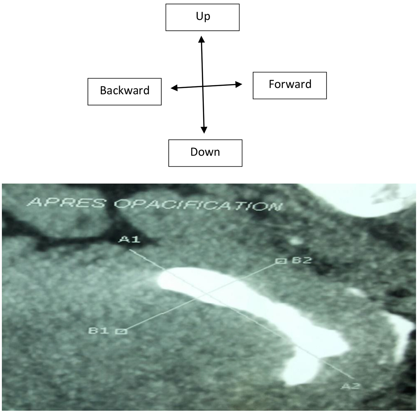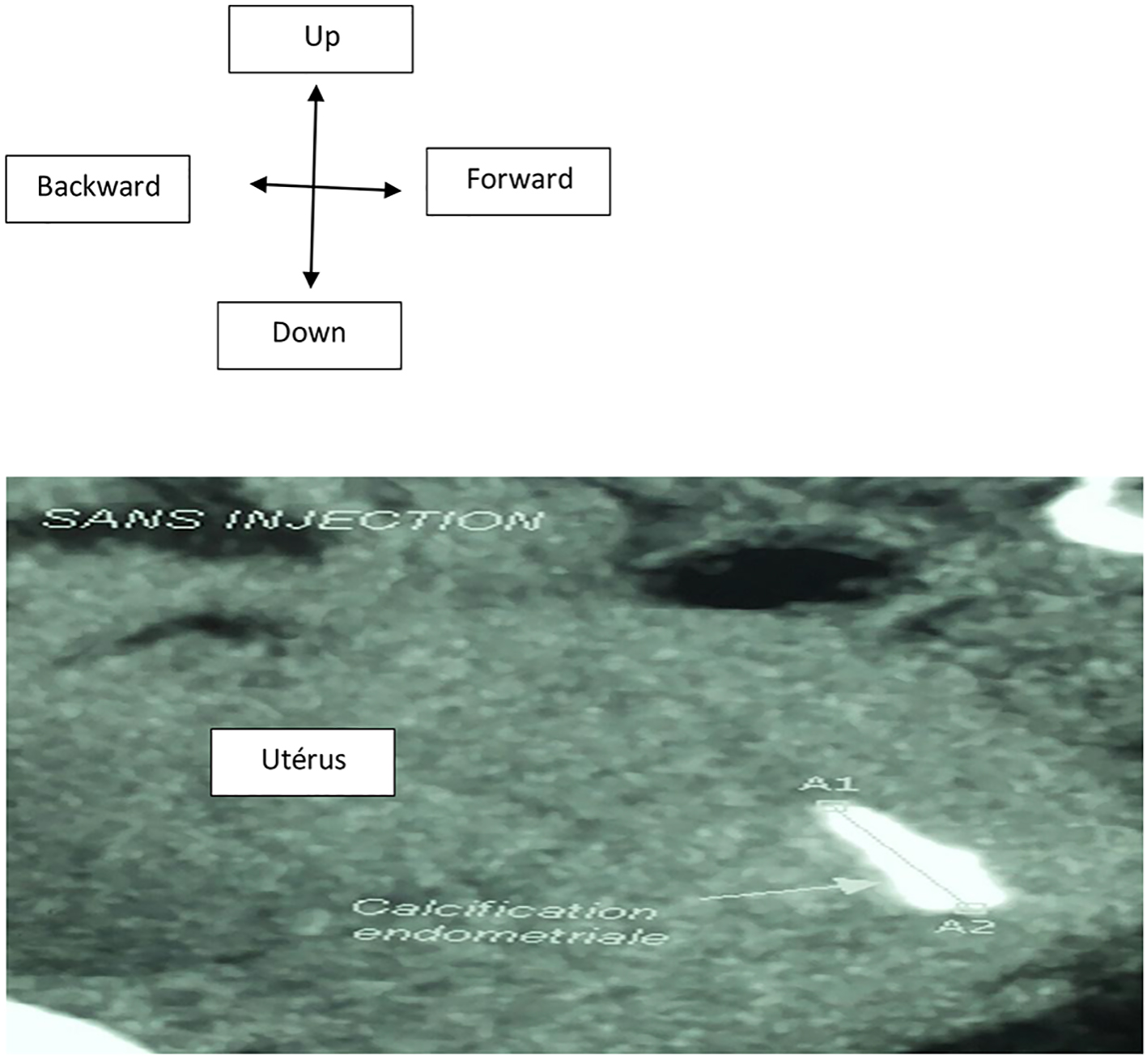
Figure 1. Hysteroscanner axial cup: before injection of the contrast agent, spontaneous hyperdensity within the endometrium about 22.3 mm diameter.
| Journal of Clinical Gynecology and Obstetrics, ISSN 1927-1271 print, 1927-128X online, Open Access |
| Article copyright, the authors; Journal compilation copyright, J Clin Gynecol Obstet and Elmer Press Inc |
| Journal website http://www.jcgo.org |
Case Report
Volume 5, Number 1, March 2016, pages 45-49
Endoscopy Management of Endometrial Ossification Associated With Secondary Infertility: A Case Report and Review of Literature
Figures