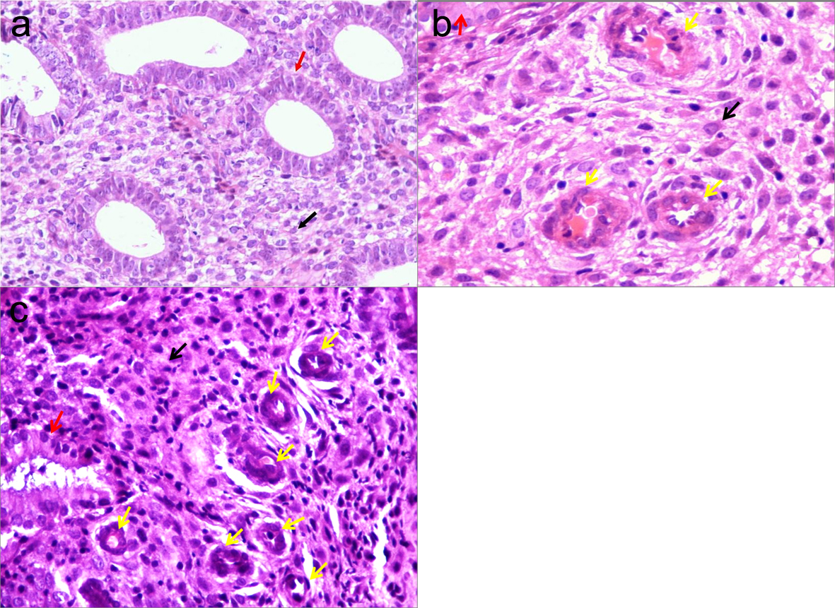
Figure 1. Photomicrograph showing poor (a), intermediate (b) and good (c) endometrial vascularity (400 X). Black arrow: stroma. Red arrow: endometrial glands. Yellow arrow: blood vessels. Flowchart of study subjects.
| Journal of Clinical Gynecology and Obstetrics, ISSN 1927-1271 print, 1927-128X online, Open Access |
| Article copyright, the authors; Journal compilation copyright, J Clin Gynecol Obstet and Elmer Press Inc |
| Journal website http://www.jcgo.org |
Original Article
Volume 2, Number 2, September 2013, pages 76-80
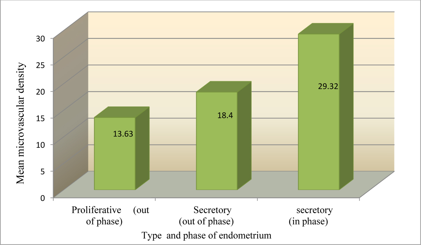
Microvascular Density as a Parameter of Endometrial Assessment in Infertile Women
Figures