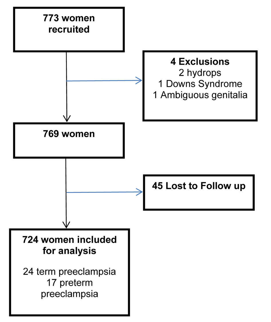
Figure 1. Flow chart demonstrating included women.
| Journal of Clinical Gynecology and Obstetrics, ISSN 1927-1271 print, 1927-128X online, Open Access |
| Article copyright, the authors; Journal compilation copyright, J Clin Gynecol Obstet and Elmer Press Inc |
| Journal website http://www.jcgo.org |
Original Article
Volume 3, Number 1, February 2014, pages 22-29
Improved Early Prediction of Preterm Pre-Eclampsia by Combining Second Trimester Maternal Serum Alpha-Fetoprotein and Uterine Artery Doppler
Figures
Tables
| Characteristic | Number n (%) n = 724 |
|---|---|
| Race | |
| Caucasian | 383 (52.9) |
| Afro-Caribbean | 289 (39.9) |
| Asian | 43 (5.9) |
| Oriental | 7 (0.9) |
| Nulliparous | 354 (48.8) |
| Smoker | 166 (22.9) |
| Previous severe pre-eclampsia | 21 (2.9) |
| Previous babies < 2,500g after 37 weeks | 11 (1.5) |
| Previous premature delivery | 12 (1.6) |
| Method | Sens(%) (True +ve/Total +ve) | Spec (%) (True -ve/True -ve +False +ve) | PPV (%) | OR (CI) | LR | P value+ | |
|---|---|---|---|---|---|---|---|
| PE: pre-eclampsia; pret PE: pre-eclampsia requiring delivery before 37 weeks gestation; Sens: sensitivity; Spec: specificity; PPV: positive predictive value; OR: odds ratio; CI: confidence intervals; LR: positive likelihood ratio. *MSAFP ≥ 2.0 MoM; ** Bilateral notches/mean RI ≥ 0.67; † Bilateral notches/mean RI ≥ 0.55 and unilateral notches/mean RI ≥ 0.65 and MSAFP > 1.2 MoM; ‡ Bilateral notches/mean RI ≥ 0.75 and unilateral notches/mean RI ≥ 0.65; # MSAFP ≥ 2.6 MoM; Φ Bilateral notches/mean RI ≥ 0.55 and unilateral notches/mean RI ≥ 0.65 combined with MSAFP ≥ 1.6 MoM. | |||||||
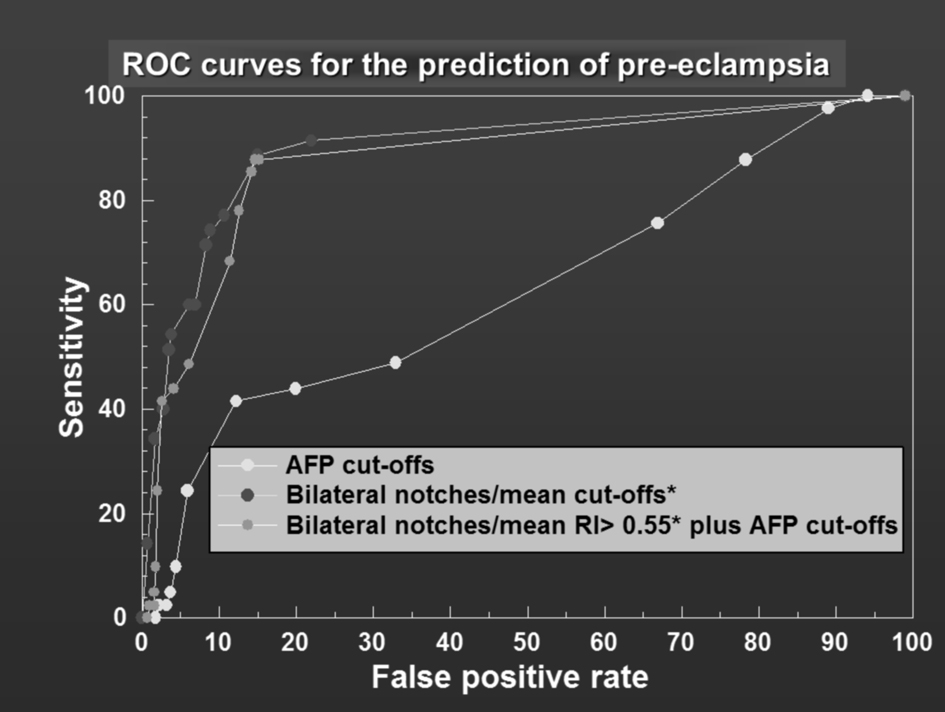
| PE | MSAFP* | 24.4 (10/41) | 94.1 (643/683) | 20.0 | 5.185 (2.4 - 11.3) | 4.1 | |
| Doppler** | 60.9 (25/41) | 92.9 (635/683) | 38.5 | 8.6 | |||
| Doppler and MSAFP† | 48.8 (20/41) | 93.9 (641/683) | 32.3 | 14.5 (7.3 - 28.9) | 8 | ||
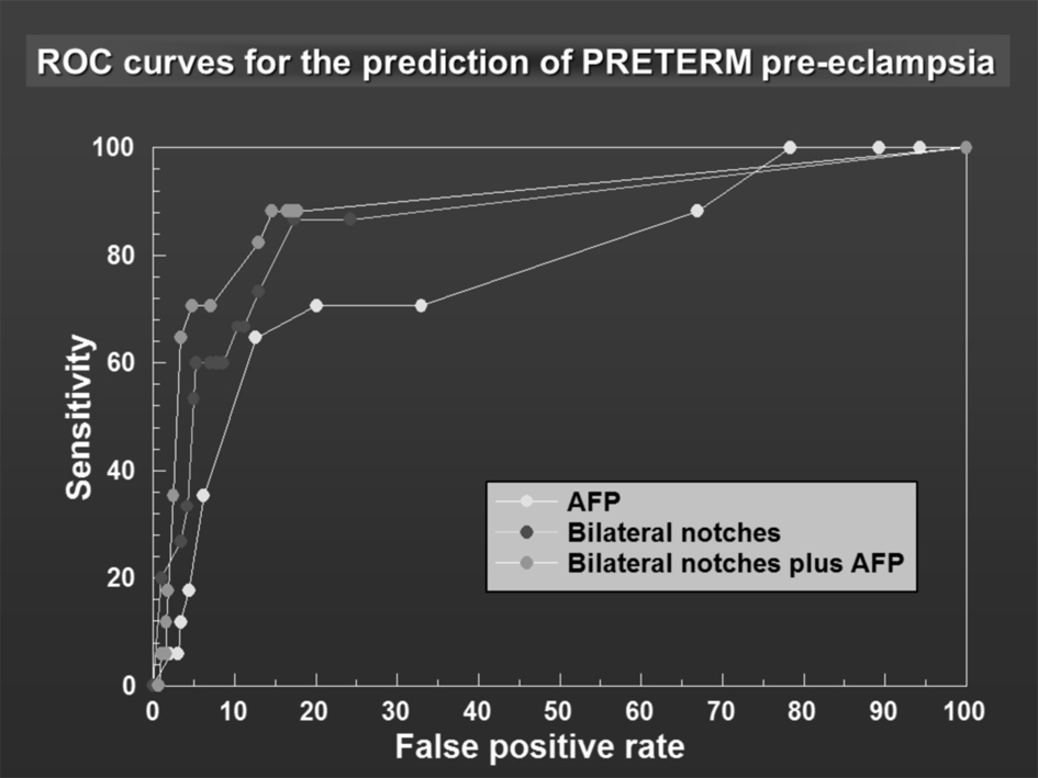
| Pret PE | MSAFP# | 5.9 (1/17) | 96.9 (685/707) | 4.3 | 9.1 (0.2 - 15.3) | 1.9 | |
| Doppler‡ | 29.4 (5/17) | 97.0 (686/707) | 19.2 | 9.8 | < 0.02 | ||
| Doppler and MSAFPφ | 64.7 (11/17) | 96.6 (683/707) | 31.4 | 52.2 (17.1 - 152.8) | 19.0 | ||
| Study | Sample | Results |
|---|---|---|
| Janiaux 1996 [16] | 41 women with abnormal UADs at 20 - 24 weeks and measured AFP | PPV 47% |
| Chung 2000 [17] | 179 women with MSAFP > 2.5MOMs and abnormal UADs between 26 - 28 weeks | PPV 14.5 % MSAFP alone |
| Konchak 1995 [18] | 103 women with unexplained increase AFP had UAD measured between 17 - 22 weeks | Elevated uterine artery RI associated with an increased RR of preeclampsia (RR 41.82, 95% CI 5.36 to 326) |
| Audibert 2005 [7] | 2,615 women had MSAFP and UAD measured in the second trimester | Elevated MSAFP with uterine artery notching had a PPV of 21% |