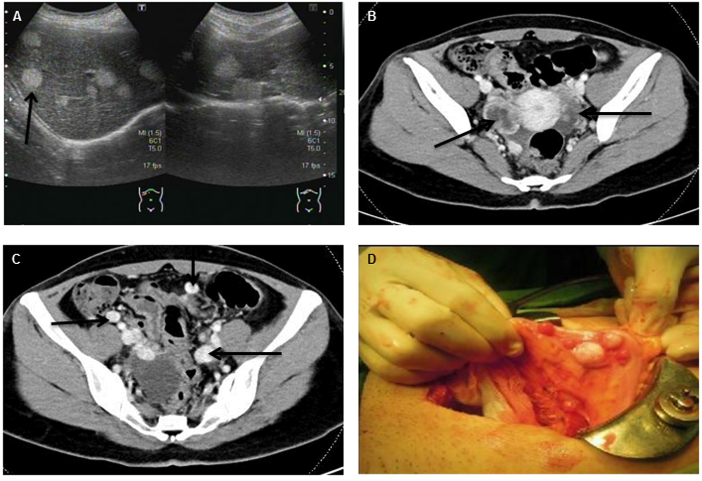
Figure 1. Abdominal ultrasonography (A) and abdominal CT with contrast enhancement (B, C) revealed multiple mixed cystic lesions over bilateral ovaries and multiple hyper-vascular tumors widespread to peritoneum, liver and lung (indicated by arrowhead). (D) Disseminated tumor seeding to peritoneum and colon (peritoneal strumosis) was demonstrated during the operation.
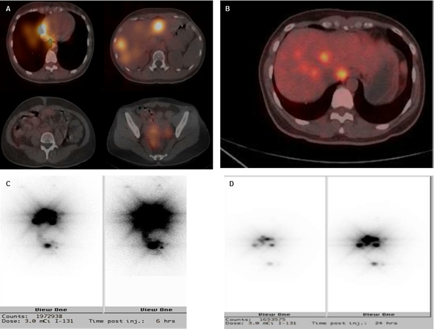
Figure 3. Imaging studies of SPECT (A), PET-FDG (B) and I131 (3 mCi) pre-treatment scan of 6 h (C) and 24 h (D). There were multiple sites of intense radiotracer uptake accumulation over the right lower lung, liver and pelvis. Relatively less intense uptake was seen over the left lower lung, right peritoneal cavity and uterine adnexa.
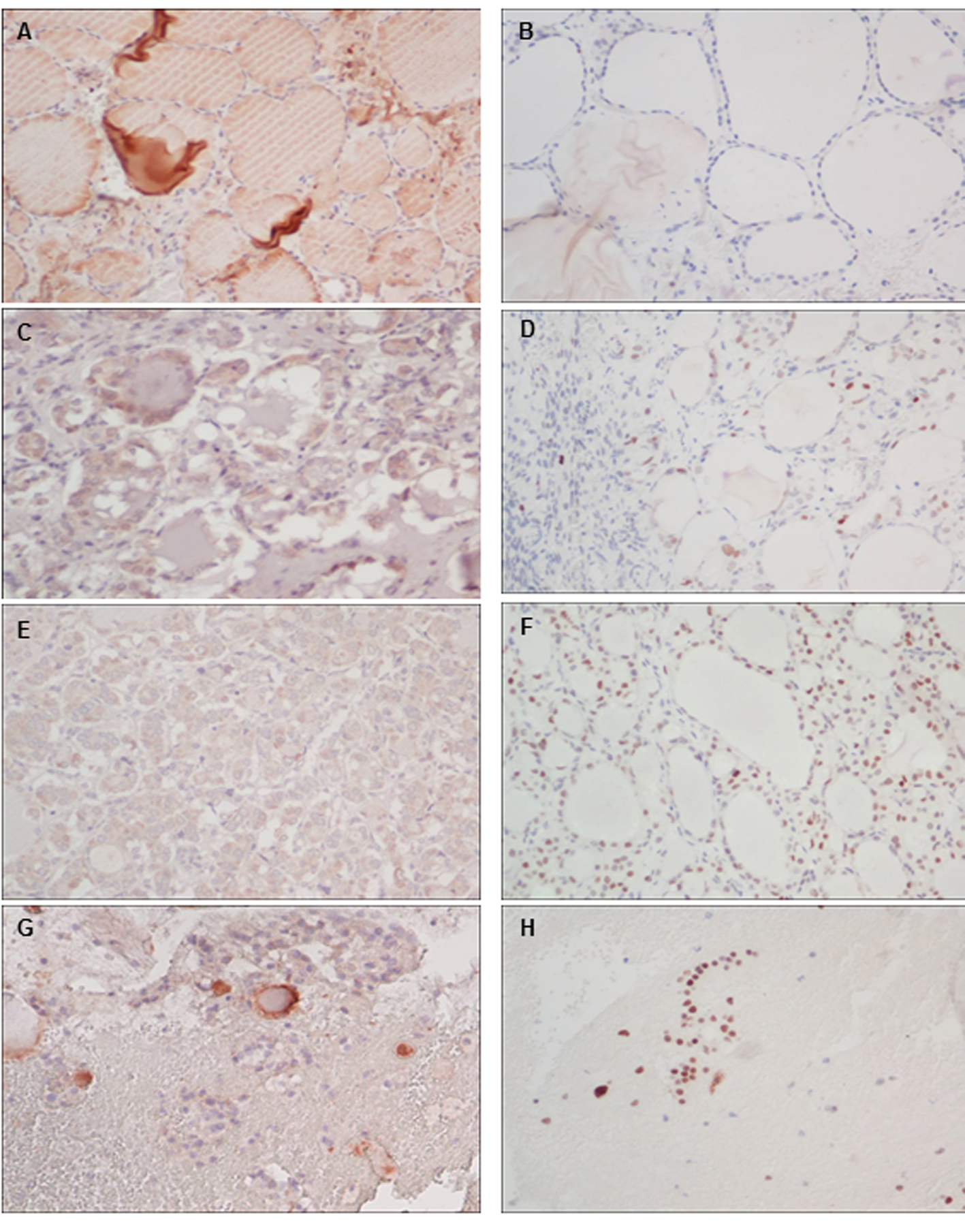
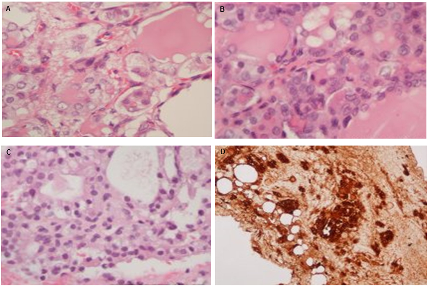
Figure 4. Immunohistochemistry (× 200) of the expressions of VEGF (left panel) and p53 (right panel) for normal thyroid (A, B), excised ovary(C, D), peritoneal (E, F) and liver seedings (G, H).