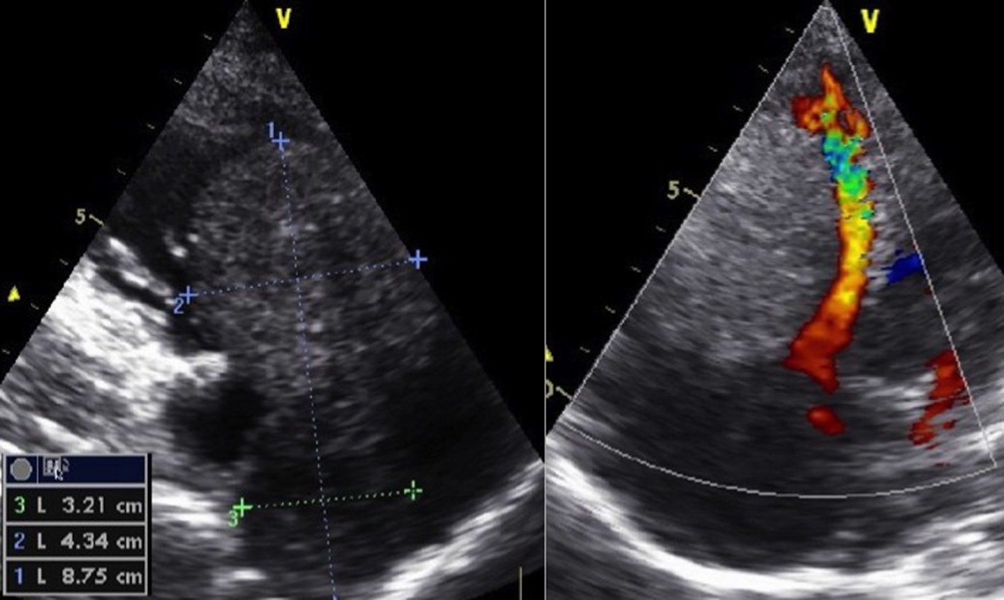
Figure 1. Modified five-chamber view of intracardiac tumor in the right ventricle.
| Journal of Clinical Gynecology and Obstetrics, ISSN 1927-1271 print, 1927-128X online, Open Access |
| Article copyright, the authors; Journal compilation copyright, J Clin Gynecol Obstet and Elmer Press Inc |
| Journal website http://www.jcgo.org |
Case Report
Volume 4, Number 1, March 2015, pages 179-183
Successful Surgical Removal of Symptomatic Intravenous Leiomyomatosis With Intracardiac Extension Completely Tamponating the Entire Right Atrium and Ventricle
Figures
Table
| Differential diagnosis | Diagnostic characteristics | Literature |
|---|---|---|
| Primary cardiac tumors, e.g. atrial myxoma | Typically limited to the heart chambers and not affecting IVC [16, 20], often attached to interatrial septum [21, 22] | Peng et al [20] Bender et al [21] Harris et al [16] Cleveland et al [22] |
| Tumor thrombus (bland or origin of other intraabdominal tumors) | No enhancement after contrast fluid in CT scan [20, 21] or MRI [23], no Doppler signal in US [23] | Bender et al [21] Peng et al [20] Fasih et al [23] |
| IVL | Women with history of uterine myoma [9, 24] Not adhesive with wall of vessels [9, 20] If there is a mass in heart normally with continuation in IVC [9] Often spindle-shaped masses, regular margin and clear boundary [20] | Xu et al [9] Peng et al [20] Liu et al [24] |
| Leiomyosarcoma | Adherent to IVC [16], arise from the wall of IVC, typical heterogeneous enhancement in contrast-enhanced MRI venography, often necrosis within the tumor [25] Only proven by histology [16] Can occur also in men [25] | Harris et al [16] Huang et al [25] |