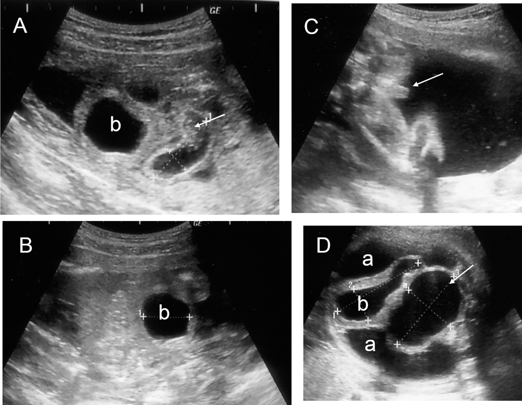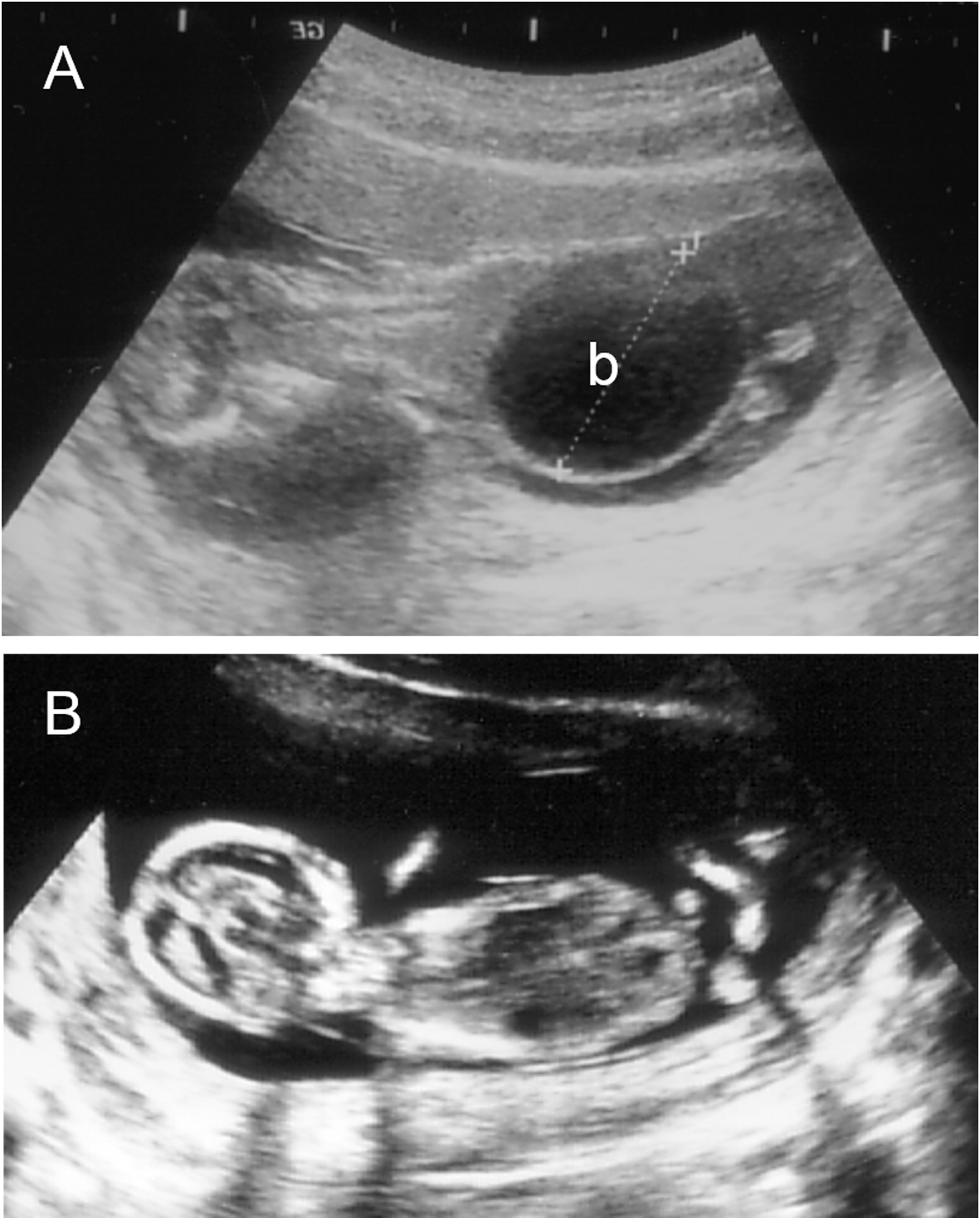
Figure 1. Longitudinal image of the fetus before (A) and after (B) the placement of a vesicoamniotic shunt. (A) 14+0 weeks of gestation; (B) 14+4 weeks of gestation. b: bladder.
| Journal of Clinical Gynecology and Obstetrics, ISSN 1927-1271 print, 1927-128X online, Open Access |
| Article copyright, the authors; Journal compilation copyright, J Clin Gynecol Obstet and Elmer Press Inc |
| Journal website http://www.jcgo.org |
Case Report
Volume 3, Number 3, September 2014, pages 117-120
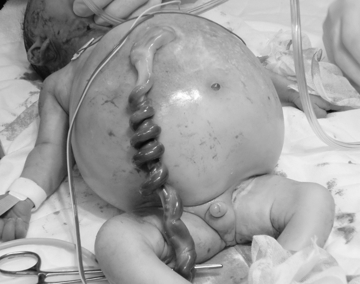
A Case of Complete Urorectal Septum Malformation Sequence: Successful Vesicoamniotic Shunting for Obstructive Uropathy
Figures