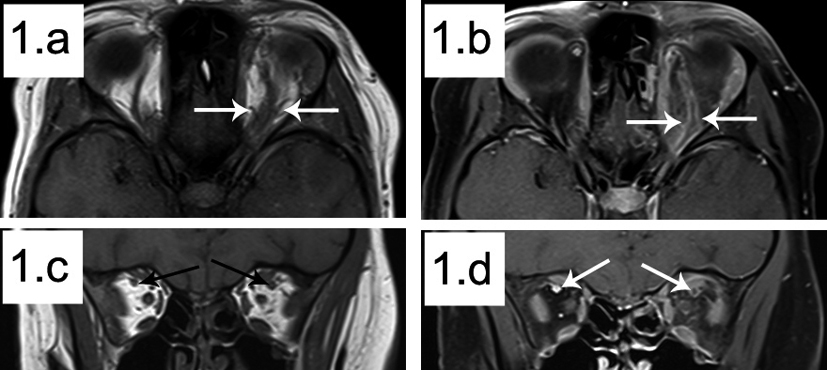
Figure 1. Coronal and Axial MRI images. a: Axial T1 showing irregularity and dilation of the left superior ophthalmic vein compared to the right (outlined by white arrows); b: Axial T1 fat sat post contrast showing thrombus in situ in the left superior ophthalmic vein (outlined by white arrows); c: Coronal T1 showing an enlarged left superior ophthalmic vein compared to the right (white arrows); d: Coronal T1 fat sat post contrast showing thrombus in situ in the left superior ophthalmic vein compared to normal contrast flow on the right (white arrows).