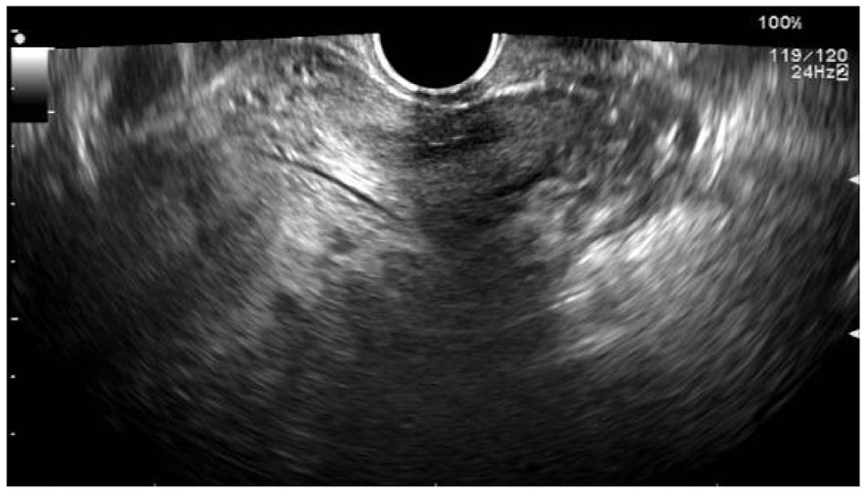
Figure 1. Transvaginal ultrasonography of the pelvic region. A 15 cm mass developed from the uterus is shown. There is no evidence of ascites or necrosis. Neither ovary is detected
| Journal of Clinical Gynecology and Obstetrics, ISSN 1927-1271 print, 1927-128X online, Open Access |
| Article copyright, the authors; Journal compilation copyright, J Clin Gynecol Obstet and Elmer Press Inc |
| Journal website http://www.jcgo.org |
Case Report
Volume 4, Number 3, September 2015, pages 275-278
Highly Elevated Level of Serum CA125 Produced by a Large Uterine Leiomyoma in a 20-Year-Old Woman
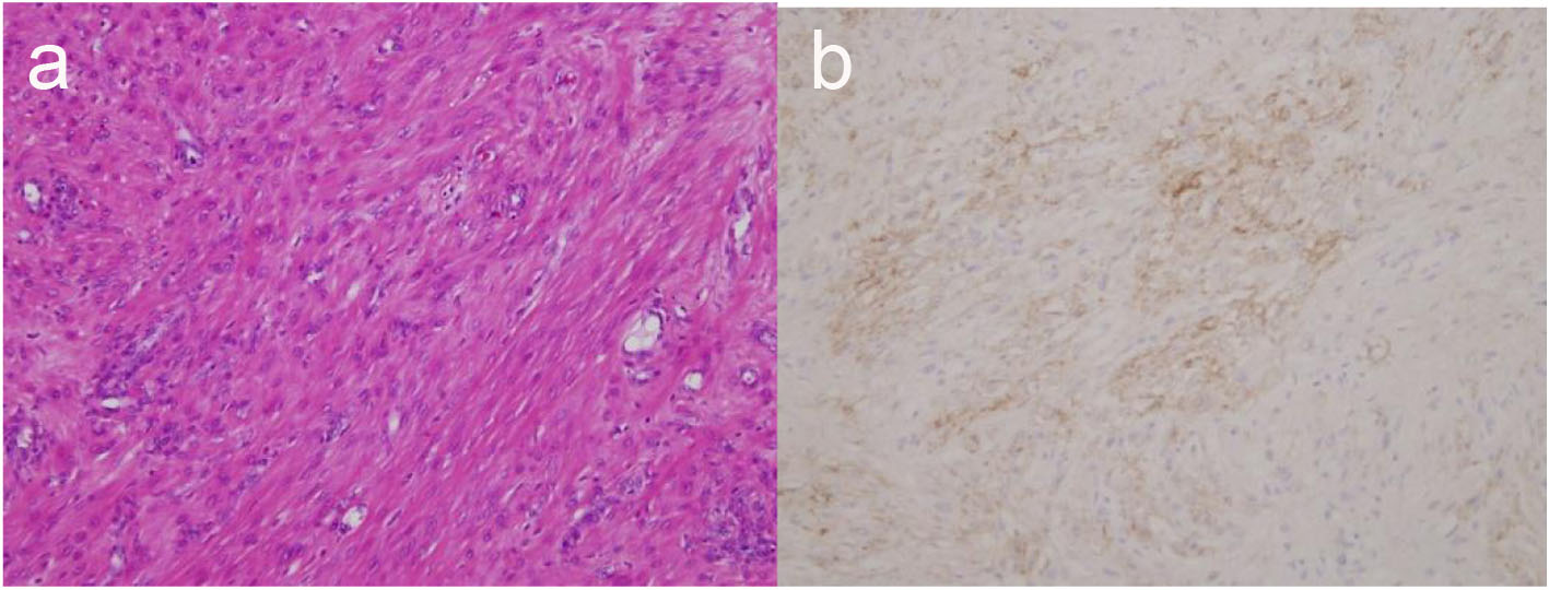
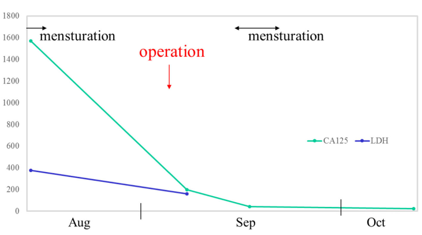
Figures