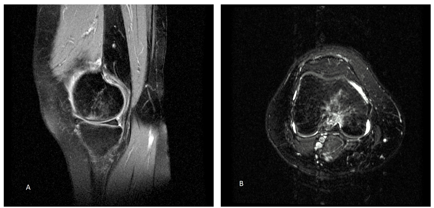
Figure 1. MRI in T2. Enhanced signal on the internal femoral condyle: (A) sagittal section and (B) axial section.
| Journal of Clinical Gynecology and Obstetrics, ISSN 1927-1271 print, 1927-128X online, Open Access |
| Article copyright, the authors; Journal compilation copyright, J Clin Gynecol Obstet and Elmer Press Inc |
| Journal website http://www.jcgo.org |
Case Report
Volume 5, Number 1, March 2016, pages 37-40
Transient Osteoporosis of the Knees, Ankles and Feet: Atypical Case in a Twin Pregnancy
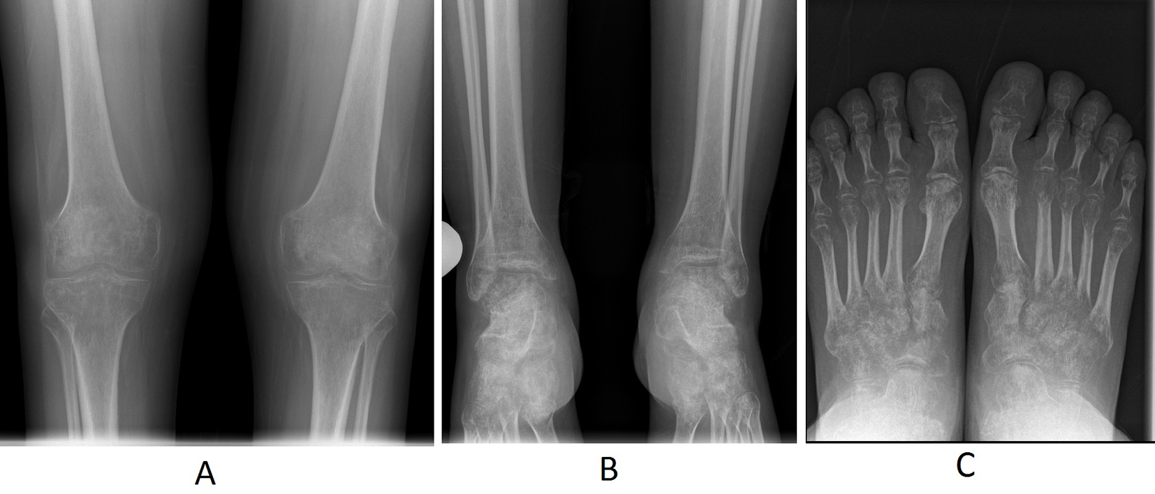
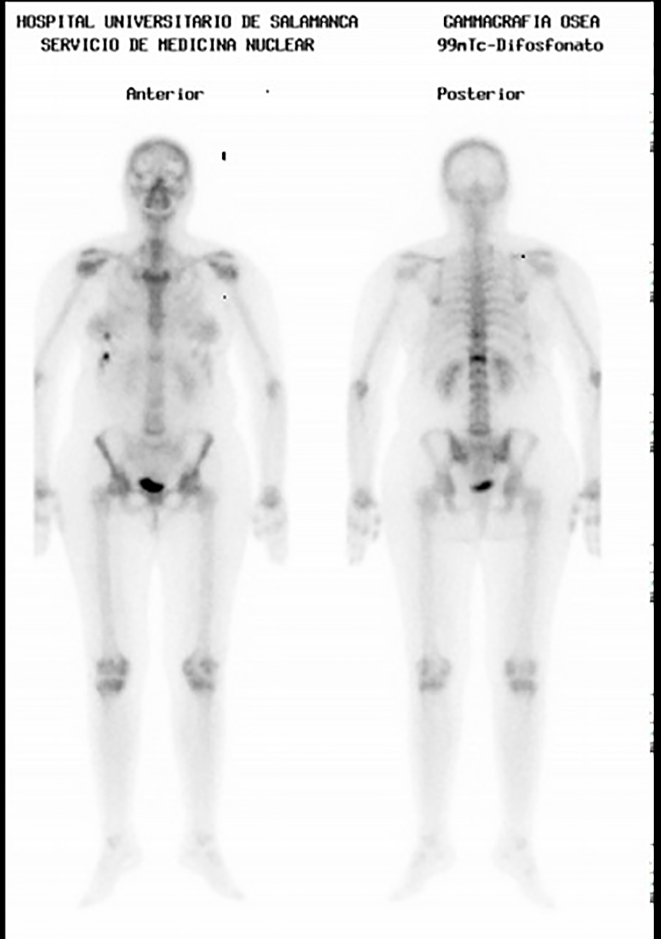
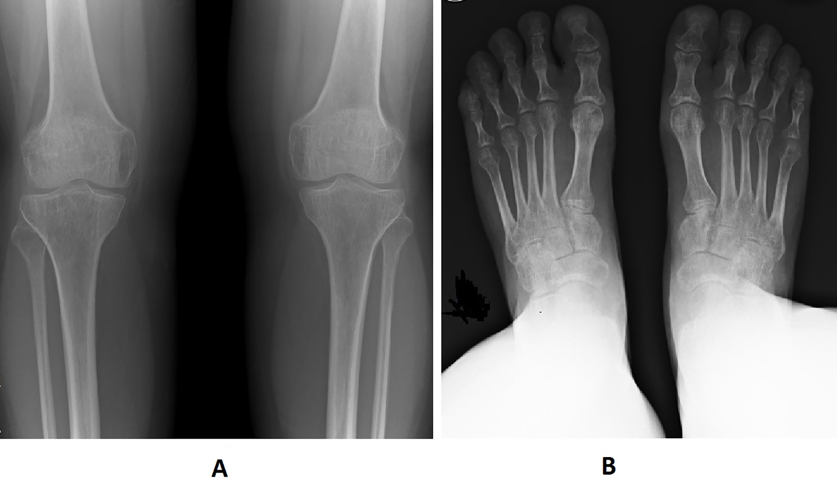
Figures