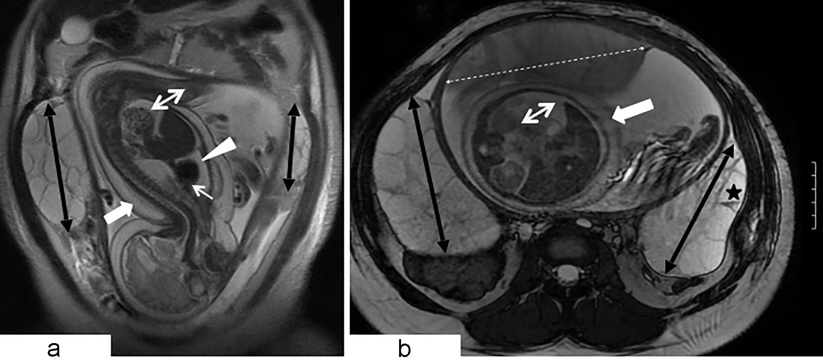
Figure 2. Coronal (a) and axial (b) views of MRI of the abdomen and pelvis (balanced steady state free precession sequence). Large gravid uterus. Cephalic position of hydropic fetus demonstrating diffuse marked skin thickening (large white arrow), large pleural effusion (white arrowhead), moderate pericardial effusion (small white arrow), and ascites (white double arrow). Bilateral flank multiseptated ovarian masses (black double arrows) composed of thin walled cysts. A few cysts reveal small fluid debris level (black star). Thickened enlarged placenta exhibits intermediate T2 signal intensity (white dashed double headed arrow). MRI findings are consistent with bilateral large ovarian theca lutein cysts (hyperreactio luteinalis) and fetal hydrops.
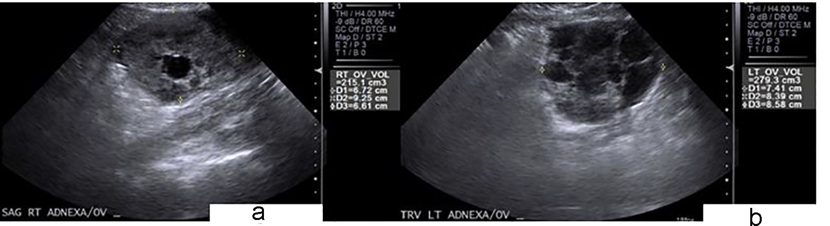
Figure 4. Transabdominal ultrasound examination performed 9 days after cesarean section. Interval decreased size of still enlarged right (a) and left (b) ovaries with decreased number of cysts. Right ovary measures 6.7 × 9.2 × 6.6 cm of 215.1 cm3 volume. Left ovary measures 7.4 × 8.3 × 8.5 cm of 279.3 cm3 volume.