Journal of Clinical Gynecology and Obstetrics, ISSN 1927-1271 print, 1927-128X online, Open Access
Article copyright, the authors; Journal compilation copyright, J Clin Gynecol Obstet and Elmer Press Inc
Journal website http://www.jcgo.org

Case Report

Volume 6, Number 2, June 2017, pages 45-48


Bilateral Thyroid-Type Struma Ovarii in a Postmenopausal Patient: A Case Report and Review of Literature

Figures

Figure 1.
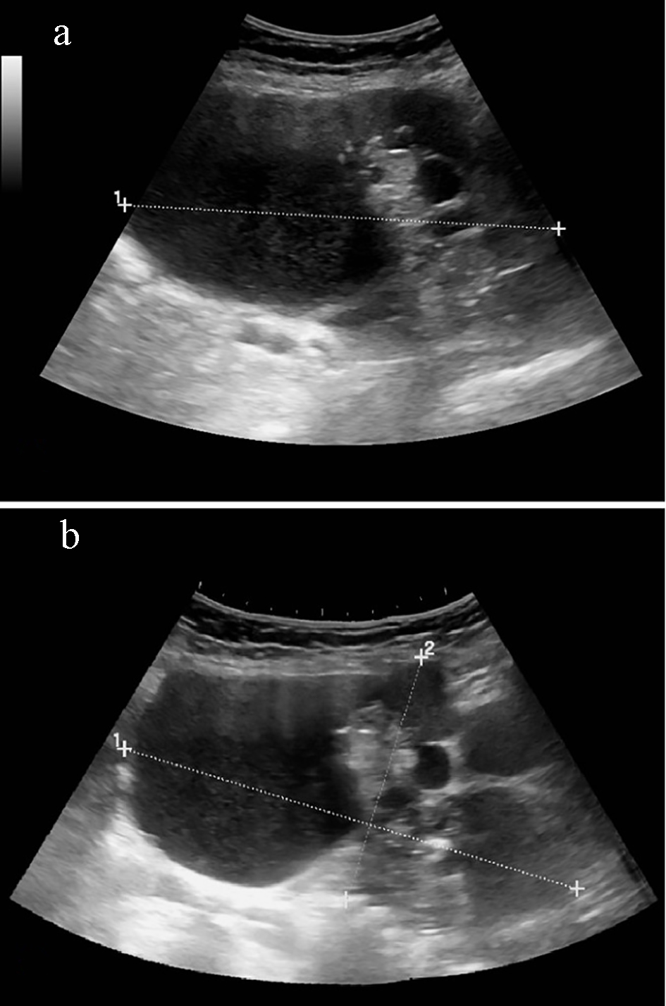
Figure 1. (a) Complex heterogenous pelvic mass with predominantly cystic and some solid echogenic areas measuring 18.68 × 13.72 × 9.76 cm (transverse view). (b) Mass involves both right and left adnexal regions and extends superiorly into the abdominal cavity (sagittal view).
Figure 2.
Figure 2. (a) Bivalved uterus with right ovarian multiloculated cyst and solid red spongy mass. (b) Left ovarian multiloculated cyst.
Figure 3.
Figure 3. (a) Ovarian stroma with mature thyroid follicles (H&E, × 100). (b) Thyroid follicular tissue containing colloid (H&E, × 400).

Table

Table 1. Summary of Selected Features of Bilateral Struma Ovarii
 
Age at diagnosis (years)Clinical presentationOvarian size (cm)Gross appearanceHistologyCA125 (U/mL) (reference: < 35 U/mL)Thyroid hormoneReference
RightLeftPreoperativePostoperative
41Metrorrhagia, abdominal distension for 2 months7.56.5MulticysticBenign thyroid follicles19Not doneNormalNovac et al, 2008 [3]
70Progressive abdominal distension, shortness of breath for 6 months27MulticysticBenign thyroid follicles28486NormalRana et al, 2009 [4]
64Slowly developing pelvic mass, pelvic organ prolapse for several years, shortness of breath812MulticysticBenign thyroid follicles12.3Not doneNormalCurrent case