Journal of Clinical Gynecology and Obstetrics, ISSN 1927-1271 print, 1927-128X online, Open Access
Article copyright, the authors; Journal compilation copyright, J Clin Gynecol Obstet and Elmer Press Inc
Journal website http://www.jcgo.org

Case Report

Volume 6, Number 2, June 2017, pages 49-52


Serous Endometrial Intraepithelial Carcinoma: Case Report and Literature Review

Figures

Figure 1.
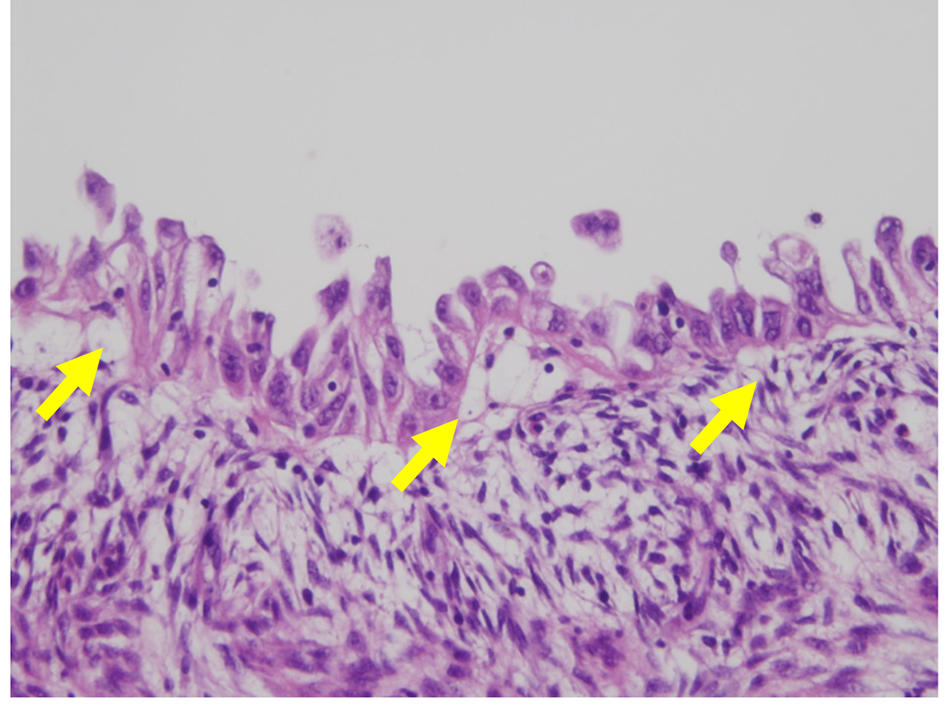
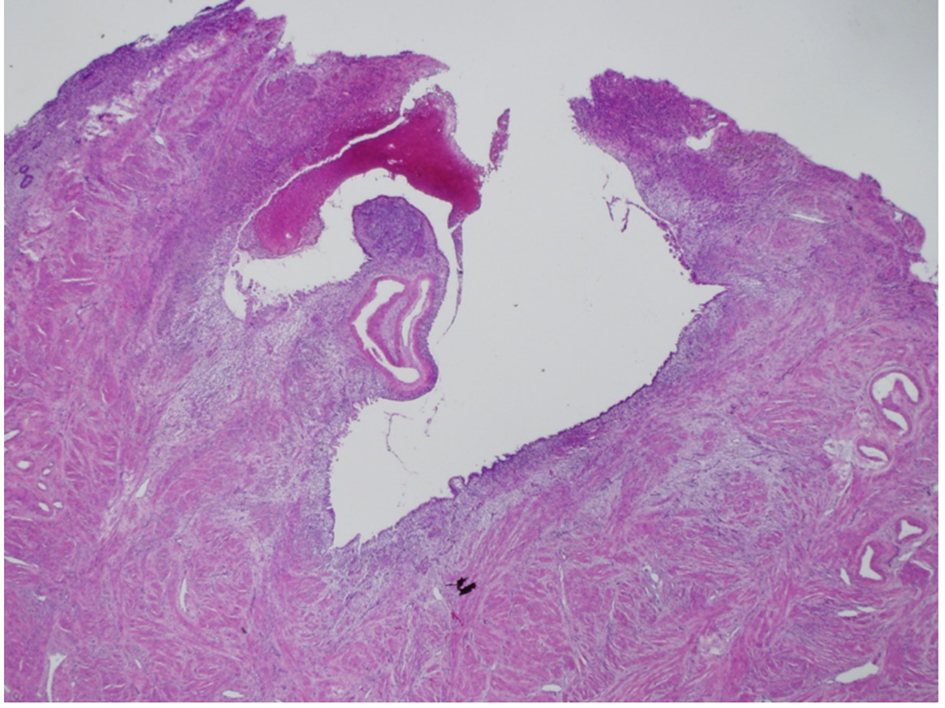
Figure 1. Histological findings of polyp from the posterior uterine wall endometrium. A cystoid tumor spanned the uterine lesion by forming confluent glands (H&E stain; original magnification × 40).
Figure 2.
Figure 2. Histological findings of the endometrium. The tumor cells formed multiple layers, and had irregular eosinophilic nucleoli, giving the nucleus a hobnail appearance (arrows). The SEIC lesion is limited to the epithelium, showing no stromal invasion. (H&E stain; original magnification × 400).
Figure 3.
Figure 3. Immunohistochemical findings for the endometrium, showing strongly positive p53 immunostaining in the nucleus of tumor cells (p53; original magnification × 400).
Figure 4.
Figure 4. Immunohistochemical findings for the endometrium. Tumor cells have lost estrogen receptor (ER) immunoreactivity (arrow) (ER; original magnification × 400).
Figure 5.
Figure 5. Immunohistochemical findings for progesterone receptor (PgR) in SEIC of the endometrium. Malignant cells have lost PgR immunoreactivity (arrow) (PgR; original magnification × 400).