Journal of Clinical Gynecology and Obstetrics, ISSN 1927-1271 print, 1927-128X online, Open Access
Article copyright, the authors; Journal compilation copyright, J Clin Gynecol Obstet and Elmer Press Inc
Journal website http://www.jcgo.org

Case Report

Volume 7, Number 1, March 2018, pages 20-22


Chemical Inflammation Associated With Adhesion Barrier Following Cesarean Section

Figures

Figure 1.
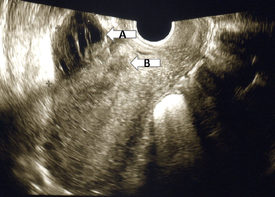
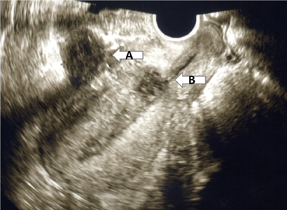
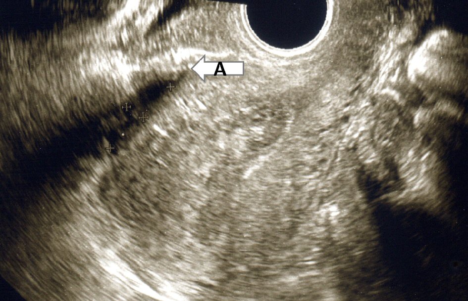
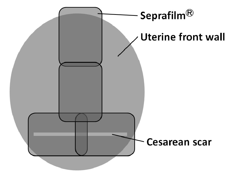
Figure 1. Four sheets of adhesion barrier (Seprafilm®) were placed in front of the uterine anterior wall.
Figure 2.
Figure 2. A low-density area of 55 × 40 mm having a high-density well-defined border indicating capsulized fluid (arrow A) was observed in front of the uterine anterior wall by ultrasonography. The position was different from that of the cesarean section scar (arrow B).
Figure 3.
Figure 3. A low-density area having a high-density well-defined border (arrow A) was observed in front of the uterine anterior wall by ultrasonography. The position was different from that of the cesarean section scar (arrow B).
Figure 4.
Figure 4. The size of low-density area having a high-density well-defined border was (arrow A) decreased significantly.