Figures
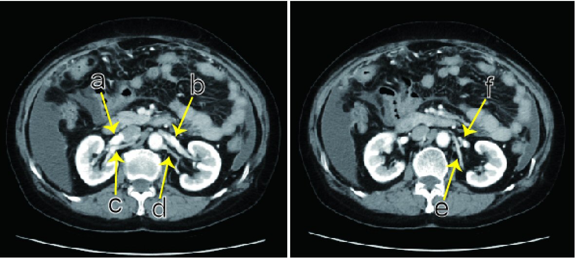
Figure 1. Abdominal contrast CT. Preoperatively, two left renal arteries are present (d, e). (a) Right renal vein. (b) The vein from the left kidney goes over an abdominal aorta, connecting at the point right before the abdominal aorta. (c) Right main renal artery. (d) Left main renal artery diverges to the apical and upper segmental arteries. (e) Left posterior segmental artery. (f) The left ovarian vein flows into the left renal vein.
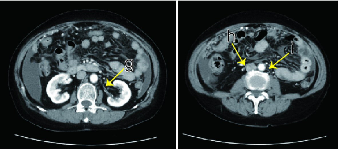
Figure 2. Abdominal contrast CT. The left middle segmental artery (g) and bilateral lower segmental artery (h, i) were not recognized preoperatively. The right lower segmental artery (h) goes over the front of the inferior vena cava.
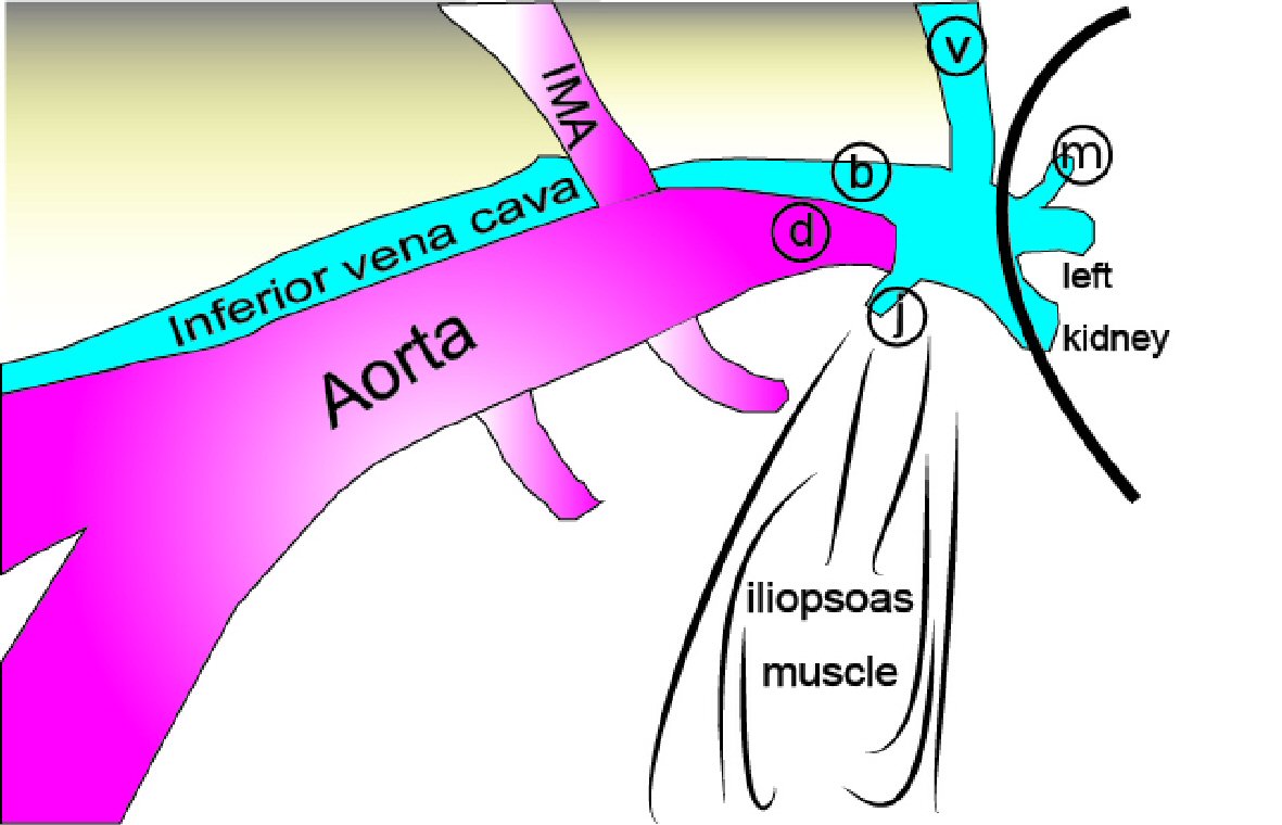
Figure 3. Normal anatomy under retroperitoneal endoscopy (left para-aortic lymphadenectomy). The left ovarian (v) and lumbar (j) veins flow into the left renal vein (b) and go over the aorta. Usually, it cannot be confirmed with the presence of fat tissues, but the left adrenal vein (m) also flows into the left renal vein. During surgery, the left ovarian vein, inferior mesenteric artery (IMA), and ureter are pushed up to the ventral side. (d) Left renal artery.
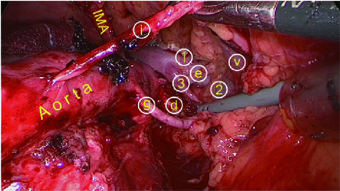
Figure 4. After left para-aortic lymphadenectomy under retroperitoneal endoscopy, three veins from the left kidney are present: (1) cephalic, (2) middle: a swollen left ovarian vein (v) flowing into the (3) caudal side (inflow lumbar vein cut). (d) Left main renal artery. (e) Left posterior segmental artery. (g) Left middle segmental artery. (i) Left lower segmental artery.
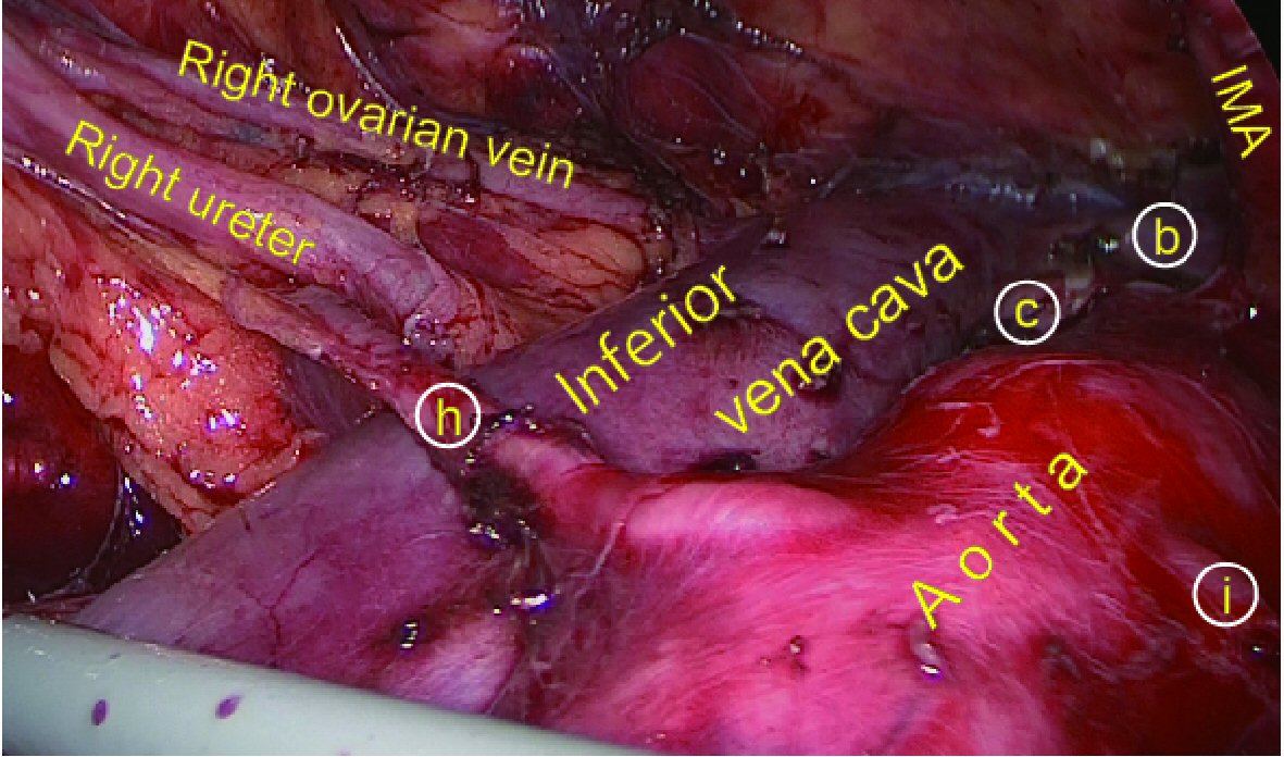
Figure 5. After right para-aortic lymphadenectomy under retroperitoneal endoscopy. (b) Left renal vein. (c) Right main renal artery. (h) Right lower segmental artery. (i) Left lower segmental artery.