Journal of Clinical Gynecology and Obstetrics, ISSN 1927-1271 print, 1927-128X online, Open Access
Article copyright, the authors; Journal compilation copyright, J Clin Gynecol Obstet and Elmer Press Inc
Journal website https://www.jcgo.org

Case Report

Volume 9, Number 3, September 2020, pages 73-77


Endometrial Stromal Sarcoma in the Young

Figures

Figure 1.
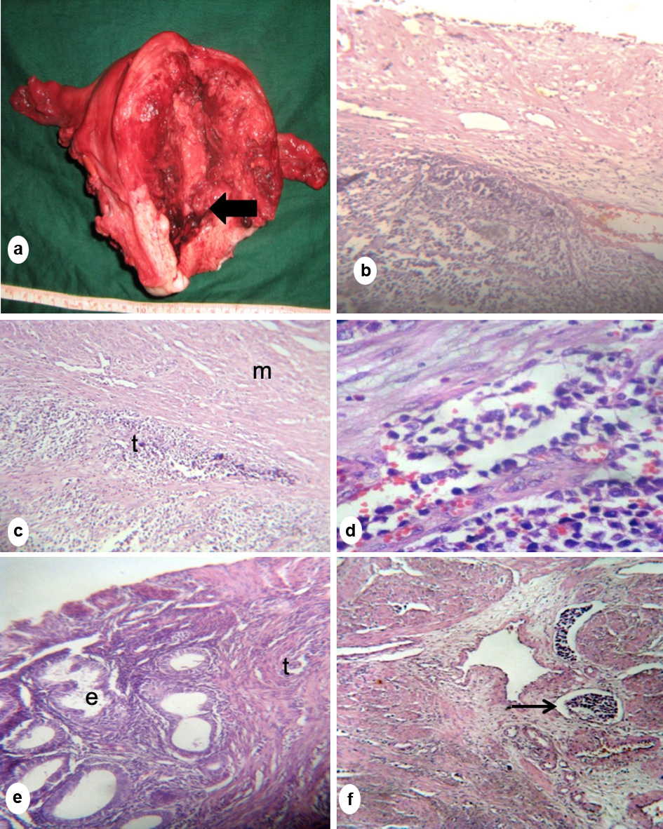
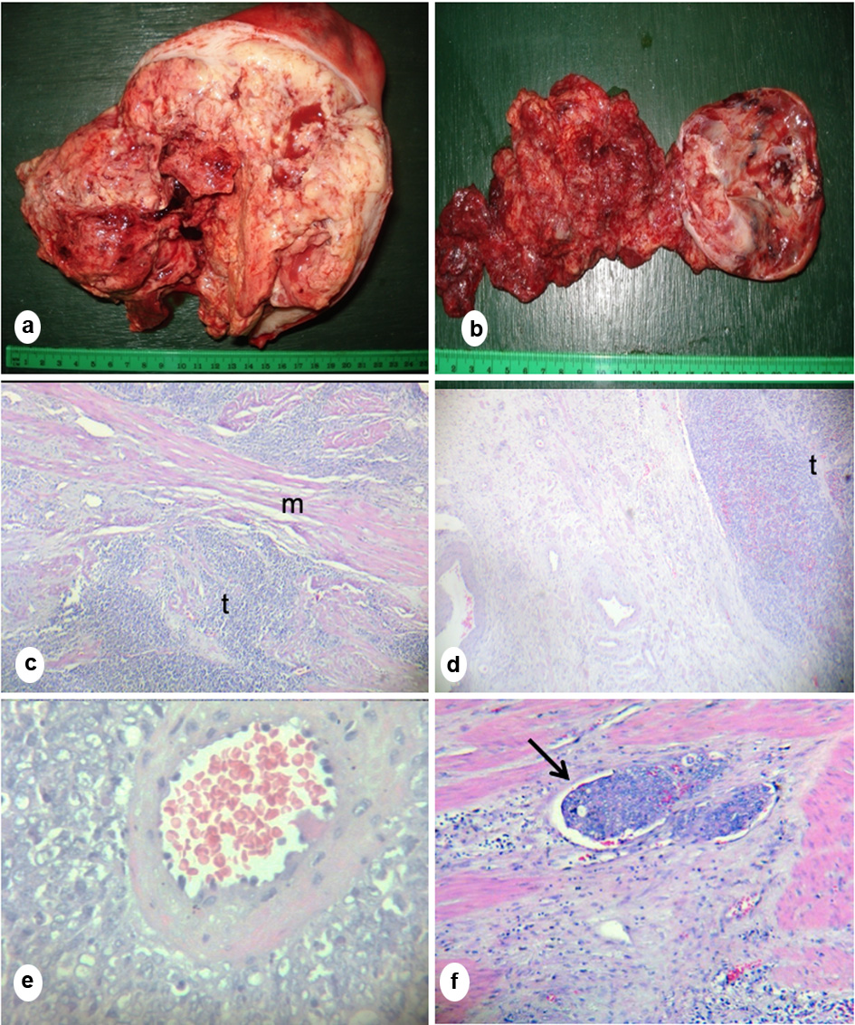
Figure 1. (a) Gross section of the uterus showed a polypoid, necrotic mass occupying the entire endometrial cavity with full thickness myometrial invasion. (b) Gross section of the right ovary showed friable necrotic mass. (c) Microscopic section of the mass showed sheaths of tumor cells (t) infiltrating smooth muscle bundles (m). (d) Microscopic section of the right ovary showed medullary tumor invasion (t). (e) Microscopic section of the tumor showing characteristic whirling of tumor cells around the spiral arteriole and (f) Lymphovascular space invasion (arrow) showing presence of tumor inside a blood vessel (c-f, hematoxylin eosin stain; c, d, f, × 20; e, × 100).
Figure 2.
Figure 2. (a) Gross section of the uterus showed a necrotic friable pedunculated mass attached to the lower uterine segment (arrow). (b) Microscopic section showing tumor infiltration extending into the posterior cervix. (c, d) Microscopic section of the uterine mass showing sheets of tumor (t) invading smooth muscle bundles of the myometrium (m), separating them sheath by sheath. (e) Endometrial glands (e) were scanty, located at the periphery of the mass and surrounded by neoplastic stromal cells (t). (f) Lymphovascular space invasion (arrow) (b-f, hematoxylin eosin stain; b, c, f, × 20; d, × 40).

Table

Table 1. Clinicopathological Profile of the Two Cases of ESS in the Young
 
Case 1Case 2
ESS: endometrial stromal sarcoma; LN: lymph node; HG-ESS: high-grade ESS; LG-ESS: low-grade ESS; EBRT: external beam radiation therapy.
Age2820
GravidityG0G0
Chief complaintVaginal bleedingVaginal bleeding
Physical examination18 × 15 cm solid abdominopelvic mass3 × 3 cm prolapsing mass at the endocervical canal
Intraoperative findingsHemoperitoneum; necrotic solid mass occupying entire endometrial cavity (10 × 8 × 2 cm); right ovary converted into a 10 × 8 × 2 cm solid mass; normal left ovary4 × 3 × 2.5 cm necrotic mass friable pedunculated mass at the isthmus; 5 × 3 × 3 cm necrotic mass friable pedunculated mass at the cervical lip; normal ovaries
LNMatted fixed pelvic LNs; 6 × 4 cm left paraaortic LNPalpable LNs
StagingIIIcIb
Histopathology resultsHG-ESSLG-ESS
Adjuvant treatmentExpired prior to adjuvant therapyHormone therapy; pelvic EBRT