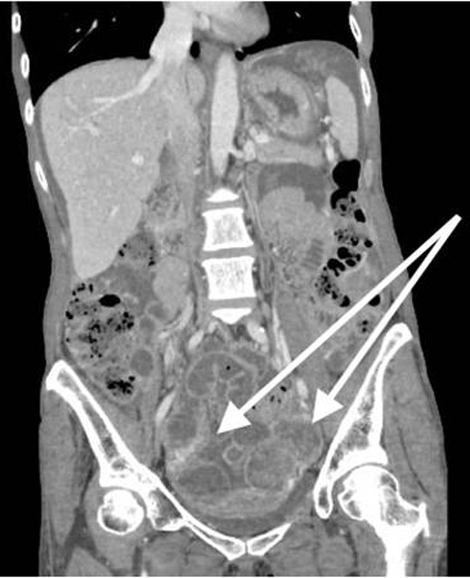
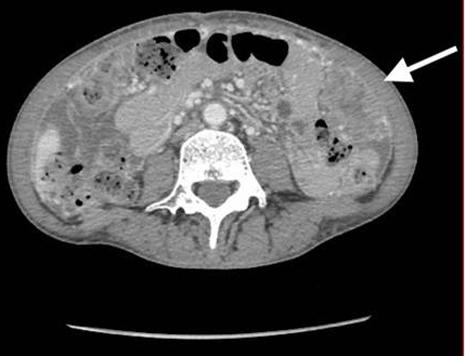
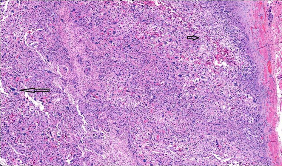
Figure 1. CT of abdomen and pelvis shows bilateral ovarian masses, and short arrow shows left ovarian mass. CT: computed tomography.
| Journal of Clinical Gynecology and Obstetrics, ISSN 1927-1271 print, 1927-128X online, Open Access |
| Article copyright, the authors; Journal compilation copyright, J Clin Gynecol Obstet and Elmer Press Inc |
| Journal website https://www.jcgo.org |
Case Report
Volume 9, Number 3, September 2020, pages 53-59
High-Grade Serous Carcinoma of Ovary With Choriocarcinomatous Differentiation: A Case Report and Review of Literature
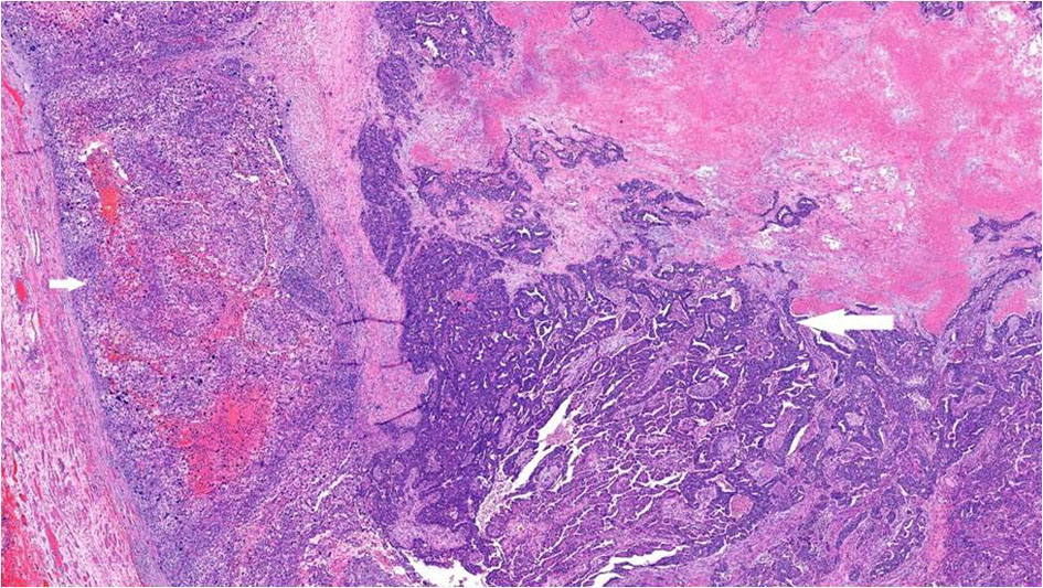
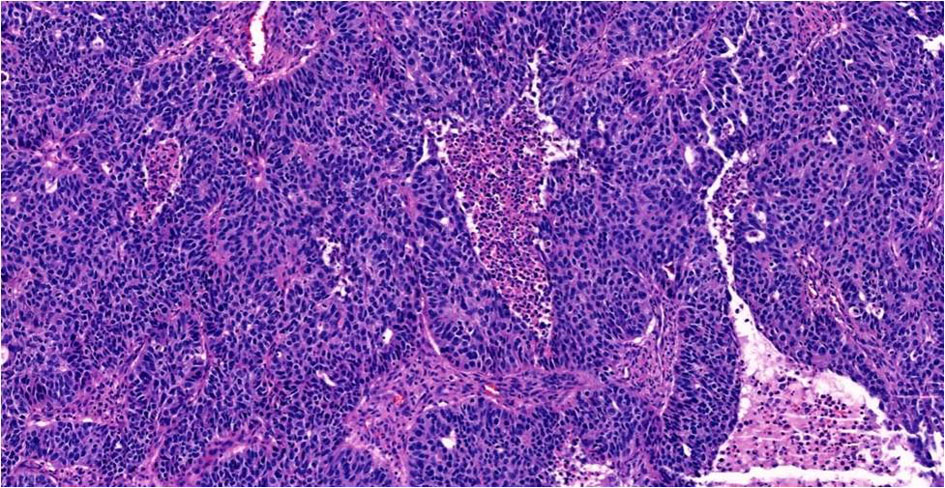
Figures
Tables
| Authors | IHC expression in choriocarcinomatous component | IHC expression in epithelial component |
|---|---|---|
| SCT: syncytiotrophoblast; CYT: cytotrophoblast; HPL: human placental lactogen; PLAP: placental alkaline phosphatase; AFP: alpha-fetoprotein; CEA: carcinoembryonic antigen; Chr: chromogranin; Syn: synaptophysin; EMA: epithelial membrane antigen; PDL-1: programmed death ligand-1; IHC: immunohistochemistry; β-hCG: beta-human chorionic gonadotrophin; N/A: not applicable. | ||
| Oliva et al [3] | Case 1: β-hCG positive in multinucleated cells; case 2: β-hCG in SCT cells | N/A |
| Ozaki et al [5] | N/A | N/A |
| Jimenez-Heffernan et al [4] | β-hCG in SCT | N/A |
| Hirabayashi et al [6] | α-hCG and β-hCG positive in SCT | CEA, CA-125, CA19-9 positive in endometrioid adenocarcinoma; Chr focal positive, Syn positive in small cell carcinoma; CA-125 positive in clear cell carcinoma |
| Oladipo et al [7] | β-hCG positive; PLAP focal positive; MNF-116 (CK) positive | N/A |
| Hafezi-Bakhtiari et al [8] | α inhibin positive; HPL positive in SCT; β-hCG positive in both CYT and SCT | EMA positive; AE1/AE3 positive; P53 positive; WT1 negative |
| Hu et al [9] | HCG positive in SCT; CYT focal positive; HPL positive in CYT | CK7 positive; CD117 negative; CD30 negative; PLAP negative; AFP negative; glypican 3 negative |
| Shanmugasundaram et al [10] | β-hCG negative | |
| Koyanagi et al [11] | β-hCG positive; AFP negative | CK7 negative; CK20 positive; CDX2 positive; ER negative; CD30 negative; PLAP negative; β-hCG negative |
| Xing et al [2] | β-hCG positive; PD-L1 positive | β-hCG negative; PD-L1 negative |
| Present case | GATA-3, PLAP, CK positive; focal p63 positive; WT1, PAX-8, ER, napsin A, AFP and CD30 negative; β-hCG: negative | P53, WT1 and PAX-8 positive in serous component |
| Author | Year | Cases | Age | Size (cm) | TZ | Histological type of associated ovarian cancer | Metastatic sites | Prognosis (follow-up) |
|---|---|---|---|---|---|---|---|---|
| R: right; L: left; mets: metastases; FU: follow-up; TZ: transition zone; Y: yes; N: no. | ||||||||
| Oliva et al [3] | 1993 | 2 | 59 | 25 | N | Case 1: undifferentiated | Lung, brain | Death in 15 months |
| 33 | 30 | Y | Case 2: mucinous cystadenoma | Liver, peritoneum | Death in 7 months | |||
| Ozaki et al [5] | 2001 | 1 | 54 | 12.4 | Mucinous cystadenoma | Myometrium, liver, lung | Death in 6 days | |
| Jimenez-Heffernan et al [4] | 2002 | 1 | 63 | 17 | Y | Mucinous cystadenocarcinoma | None | No recurrence after 7 months of FU |
| Hirabayashi et al [6] | 2006 | 1 | 50 | 12.8 | Y | Endometrioid and small cell, focal clear cell component | Peritoneum, liver, lung | Death in 10 months |
| Oladipo et al [7] | 2007 | 1 | 60 | 16 | High-grade mixed (Adeno and squamous) Carcinoma | Lung, brain | Died of brain mets, over a year | |
| Hafezi-Bakhtiari et al [8] | 2010 | 1 | 65 | 5 | High-grade papillary serous carcinoma | Lung | Lung mets after 9 months of FU | |
| Hu et al [9] | 2010 | 1 | 48 | R = 6 cm, L = 5 cm | N | Clear cell carcinoma | Lung, liver, abdominopelvic | Death after 11 months |
| Shanmugasundaram et al [10] | 2015 | 1 | 64 | R = 4 cm, L = 4 cm | High-grade papillary serous carcinoma | Peritoneum | Stable after 1-year FU | |
| Koyanagi et al [11] | 2016 | 1 | 29 | 20 | N | Adenocarcinoma (mucinous carcinoma) | Lung | NA |
| Xing et al [2] | 2019 | 1 | 44 | 11.5 | Mixed clear cell and endometrioid carcinoma | Lung | Alive with multiple lung mets after 8 months | |
| Present case | 2019 | 1 | 53 | 9.4 | High-grade serous carcinoma | Liver | Death in 2 months | |