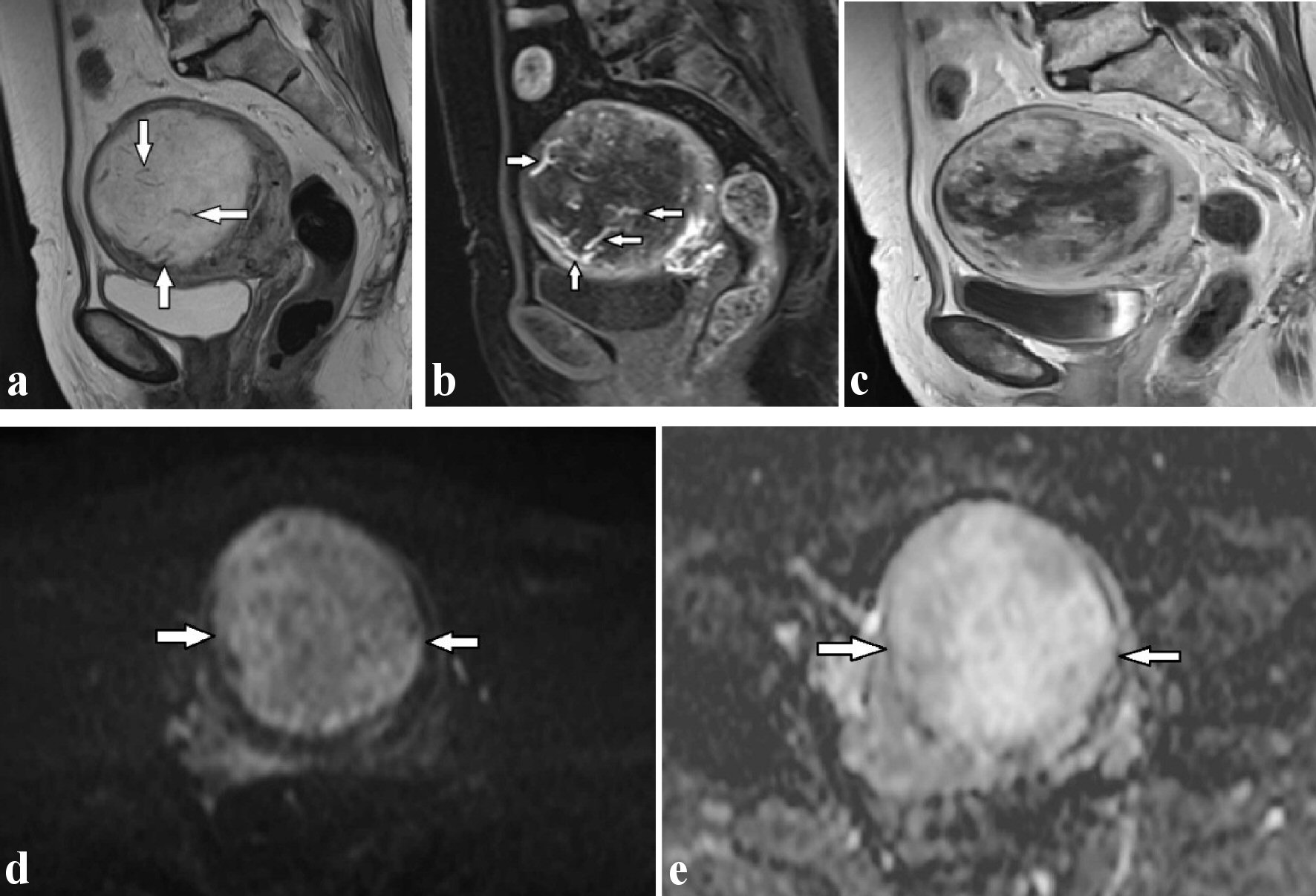
Figure 1. (a) T2-weighted sagittal image shows multiple flow-void signals (arrows) in the uterine mass revealing marked hyperintensity. (b) The flow-voids are found to correspond to dilated arteries located inside the mass in the early phase after administration of Gd-DTPA on dynamic T1-weighted images (arrows). (c) The peripheral part of the mass was gradually enhanced. Diffusion-weighted imaging (d) and apparent diffusion coefficient map (e) reveal the hyperintensity uterine mass (arrows), indicative of T2-shine through effect, which was suggestive of edematous change. Gd-DTPA: gadopentetate dimeglumine.