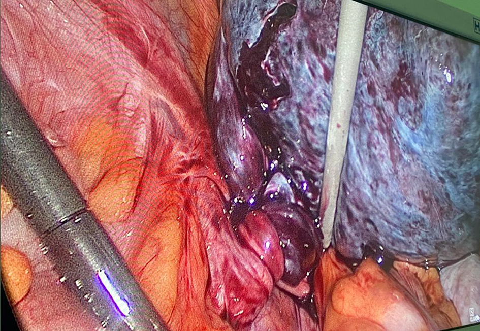
Figure 1. Intraoperative photograph showing twisted ovarian cyst.
| Journal of Clinical Gynecology and Obstetrics, ISSN 1927-1271 print, 1927-128X online, Open Access |
| Article copyright, the authors; Journal compilation copyright, J Clin Gynecol Obstet and Elmer Press Inc |
| Journal website https://www.jcgo.org |
Case Report
Volume 9, Number 4, December 2020, pages 129-133
Rare Presentation of Fetus in Fetu - Laparoscopic Approach: A Case Report
Figures
Table
| Author, year | Age | Sex | Site | Size (cm) | Symptoms | Management | Postoperative test |
|---|---|---|---|---|---|---|---|
| Largest dimension of the mass has been calculated as the size. Immunohistochemistry was not performed in any of the cases of adult FIF described in literature. Preoperative CT was the diagnostic modality in all cases where data were available. Only patients who had a clear supporting postoperative diagnosis were included in the literature review. FIF: fetus in fetu; M: male; F: female; RP: retroperitoneal; HPE: histopathological examination; CECT: contrast-enhanced computed tomography; NA: data not available. | |||||||
| Dagradi et al, 1992 [4] | 47 | M | RP | 20 | Upper abdominal mass since birth | Surgery | HPE |
| Shrivastava et al, 1999 [3] | 27 | M | RP | NA | NA | Surgery | NA |
| Awasthi et al, 2001 [5] | 30 | M | RP | 27 | Slow growing mass from childhood | Laparotomy | NA |
| Masaad et al, 2001 [6] | 27 | M | RP with mediastinal extension | 22 | Recent dysphagia, mass from childhood | Laparotomy | HPE, chromosome analysis |
| Sharma et al, 2007 [8] | 36 | M | RP | 27 | Upper abdominal swelling, vomiting, anemia | Laparotomy | Pathological examination |
| Daga et al, 2009 [9] | 20 | M | RP | 20 | Upper abdominal swelling from birth, acute pain | Laparotomy | HPE |
| Murtaza et al, 2010 [7] | 30 | F | RP | 18 | Chronic abdominal lump | Laparotomy | X-ray, HPE |
| Kumar et al, 2019 [2] | 17 | F | RP | 30 | Chronic abdominal lump | Laparotomy | CECT |