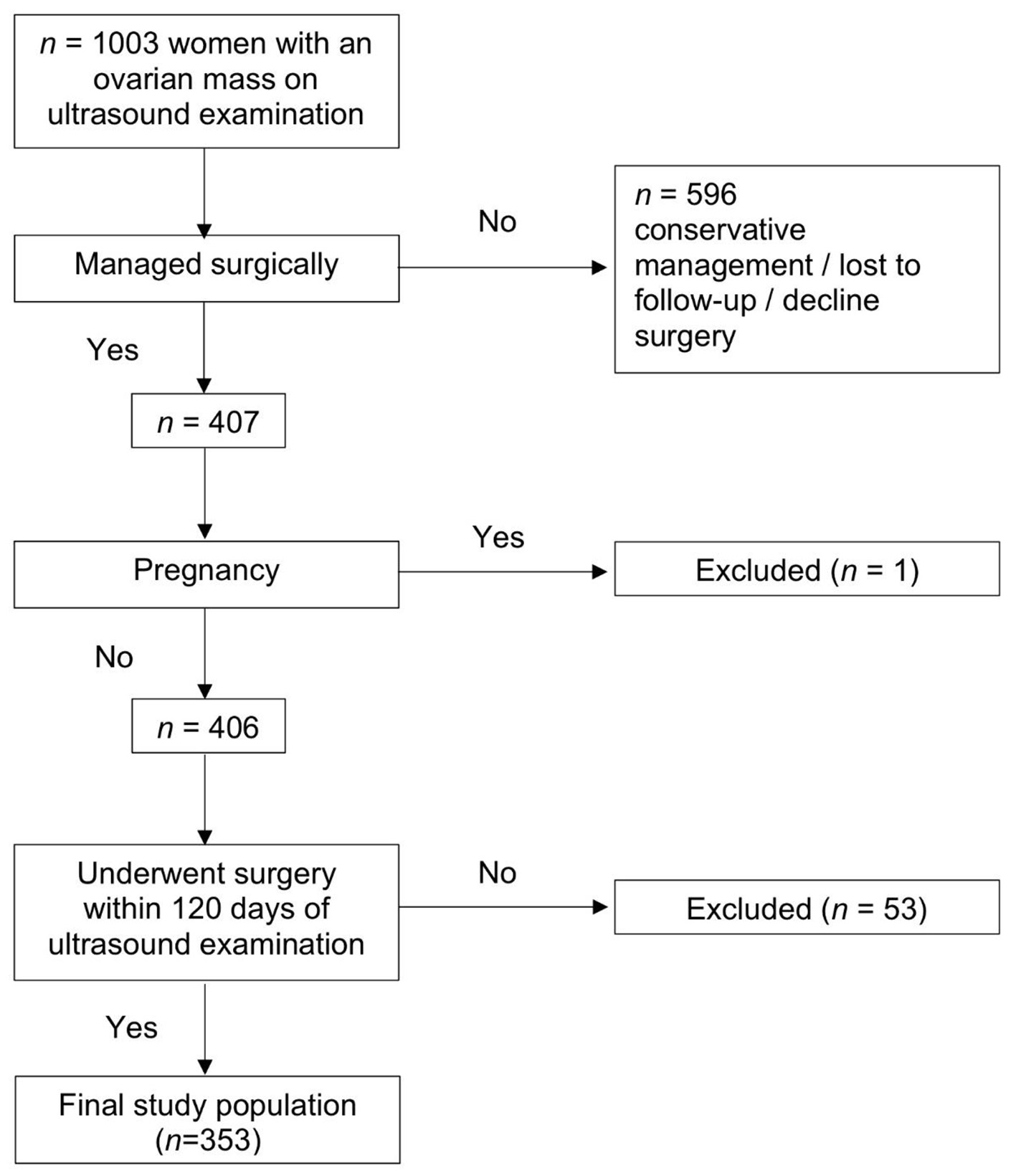
Figure 1. Flowchart of patient numbers.
| Journal of Clinical Gynecology and Obstetrics, ISSN 1927-1271 print, 1927-128X online, Open Access |
| Article copyright, the authors; Journal compilation copyright, J Clin Gynecol Obstet and Elmer Press Inc |
| Journal website https://www.jcgo.org |
Original Article
Volume 10, Number 3, September 2021, pages 67-72
Diagnostic Performance of International Ovarian Tumor Analysis Logistic Regression Model LR2 for Adnexal Masses Classification at a Tertiary Gynecology Center in Singapore
Figures
Tables
| Histological diagnosis | n (%) |
|---|---|
| aTwo cases of mesothelial inclusion cyst, one Brenner tumor, one right ovarian leiomyoma, and one adenomatoid tumor of the ovary. bCarcinosarcoma (n = 1), leiomyosarcoma (n = 1), Mullerian adenosarcoma (n = 1). | |
| Benign | 223 (63.2) |
| Mature cystic teratoma | 56 (15.9) |
| Fibroma | 6 (1.7) |
| Endometrioma | 30 (8.5) |
| Cystadenoma (serous, mucinous, seromucinous) | 74 (21.0) |
| Cystadenofibroma (serous, mucinous, seromucinous) | 15 (4.2) |
| Hemorrhagic cyst | 5 (1.4) |
| Simple ovarian cyst | 2 (0.6) |
| Hydrosalpinx | 1 (0.3) |
| Tubo-ovarian abscess | 5 (1.4) |
| Paraovarian/paratubal cyst | 6 (1.7) |
| Functional cyst | 8 (2.3) |
| Fibrothecoma | 3 (0.8) |
| Rare benign tumorsa | 5 (1.4) |
| Peritoneal inclusion cyst | 3 (0.8) |
| Borderline | 29 (8.2) |
| Mucinous | 19 (5.4) |
| Serous | 6 (1.7) |
| Seromucinous | 3 (0.8) |
| Endometrioid | 1 (0.2) |
| Primary invasive ovarian carcinoma | 87 (24.6) |
| Epithelial carcinoma | |
| High-grade serous carcinoma | 19 (5.4) |
| Endometrioid carcinoma | 26 (7.4) |
| Clear cell carcinoma | 18 (5.1) |
| Mucinous carcinoma | 14 (4.0) |
| Low-grade serous carcinoma | 3 (0.8) |
| Germ cell tumor | 4 (1.1) |
| Immature teratoma | |
| Sex cord stromal tumor | 1 (0.3) |
| Adult granulosa cell tumor | |
| Carcinosarcoma | 2 (0.6) |
| Primary uterine carcinomab | 3 (0.8) |
| Metastatic | 11 (3.1) |
| Histological diagnosis | n (%) |
|---|---|
| IOTA: International Ovarian Tumor Analysis. | |
| Cystadenofibroma | 4 (6.7) |
| Cystadenoma | 16 (26.7) |
| Endometrioma | 7 (11.7) |
| Fibroma | 3 (5.0) |
| Fibrothecoma | 1 (1.7) |
| Functional cyst | 5 (8.3) |
| Hemorrhagic cyst | 3 (5.0) |
| Broad ligament leiomyoma | 2 (3.3) |
| Peritoneal inclusion cyst | 1 (1.7) |
| Rare benign | 1 (1.7) |
| Teratoma | 16 (26.7) |
| Tubo-ovarian abscess | 1 (1.7) |
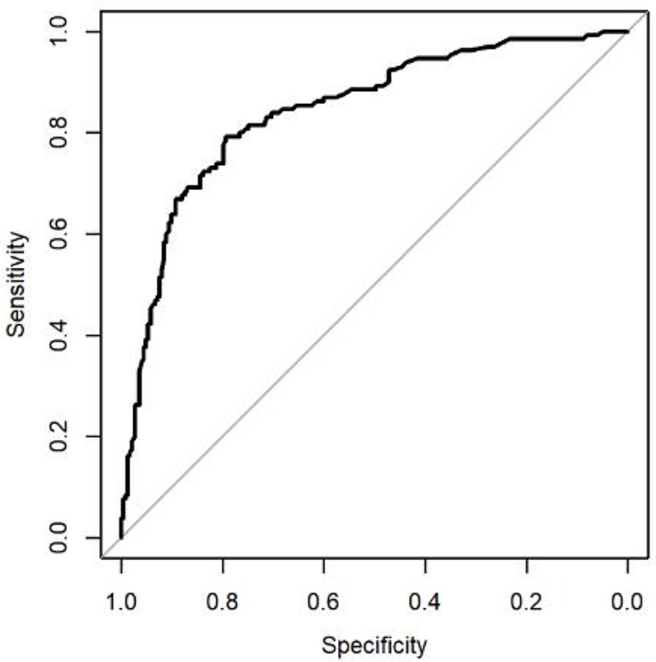
| Study | AUC (95% CI) | Sensitivity (%) | Specificity (%) | LR+ | LR- |
|---|---|---|---|---|---|
| IOTA: International Ovarian Tumor Analysis; AUC: area under receiver-operating characteristics; CI: confidence interval; LR-: negative likelihood ratio; LR+: positive likelihood ratio. | |||||
| Current study (n = 353) | 0.84 (0.80 - 0.89) | 79.2 | 79.4 | 3.84 | 0.26 |
| Original IOTA study (n = 312) [8] | 0.92 | 89.0 | 73.0 | 3.3 | 0.15 |
| Temporal validation study (n = 941) [9] | 0.92 (0.90 - 0.94) | 89.2 | 79.8 | 4.4 | 0.14 |
| External validation study (n = 997) [9] | 0.95 (0.93 - 0.96) | 91.8 | 85.6 | 6.36 | 0.10 |