Journal of Clinical Gynecology and Obstetrics, ISSN 1927-1271 print, 1927-128X online, Open Access
Article copyright, the authors; Journal compilation copyright, J Clin Gynecol Obstet and Elmer Press Inc
Journal website https://www.jcgo.org

Case Report

Volume 11, Number 1, March 2022, pages 14-18


Eclampsia in the Previable Period of 22w5d

Figures

Figure 1.
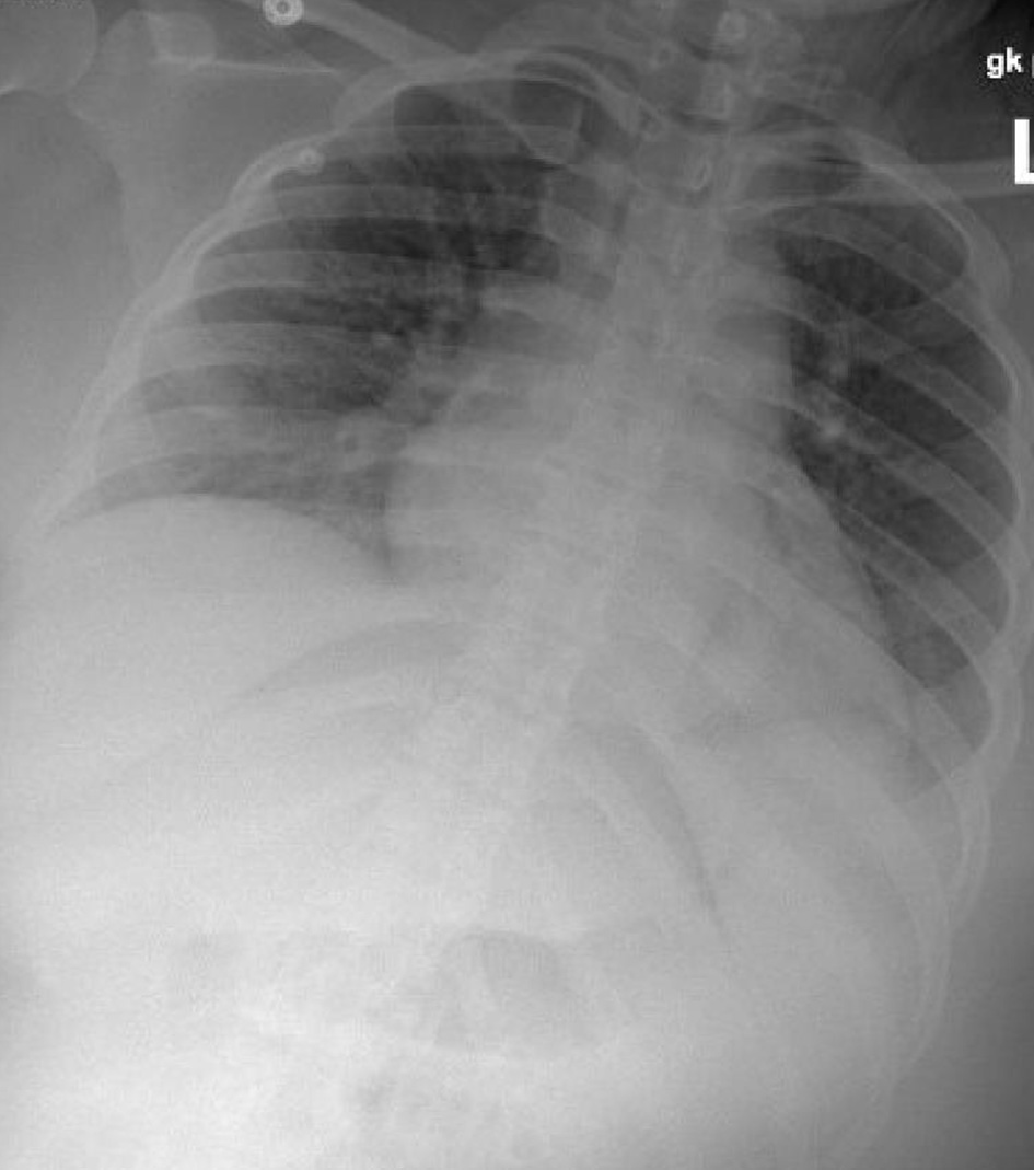
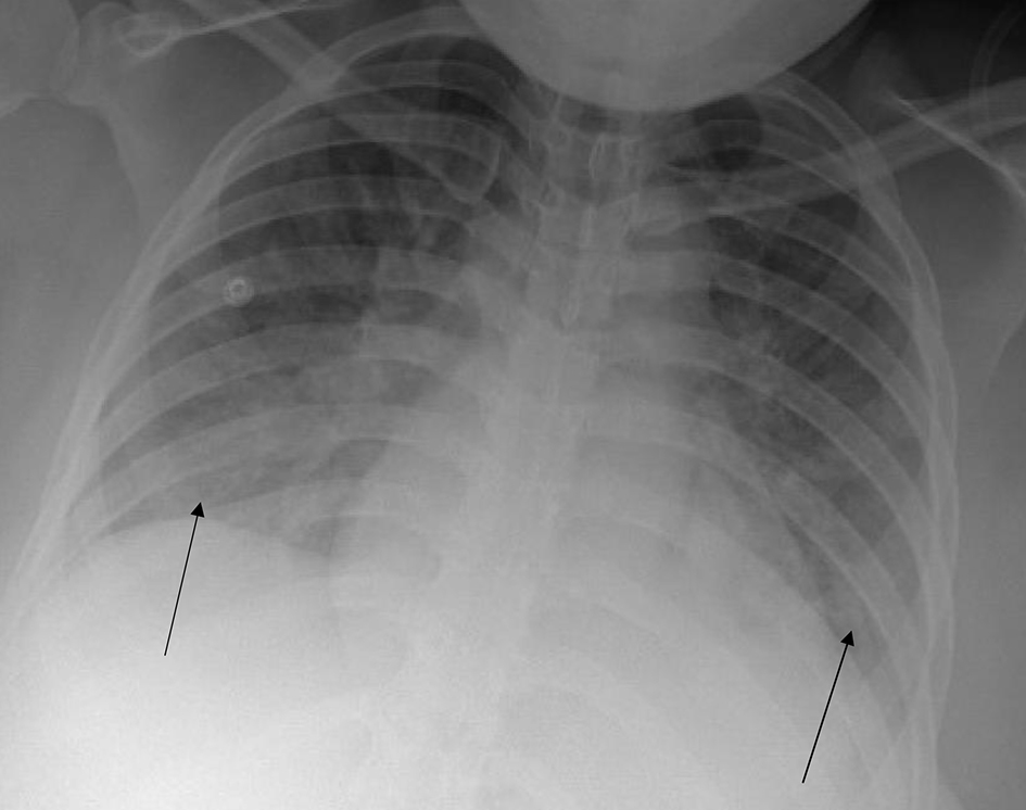
Figure 1. Chest X-ray prior to delivery. Arrows indicate diffuse pulmonary edema.
Figure 2.
Figure 2. Head computed tomography. Arrow indicates confluent white matter hypodensity within the vertex and posterior parieto-occipital region, consistent with posterior reversible encephalopathy syndrome (PRES).
Figure 3.
Figure 3. Chest X-ray post-delivery showing low lung volumes and clear lungs.
Figure 4.
Figure 4. Electrocardiogram on post-partum day 2 showing normal sinus rhythm.
Figure 5.
Figure 5. Computed tomography angiogram to rule out pulmonary embolism. No pulmonary embolism was visualized.