Journal of Clinical Gynecology and Obstetrics, ISSN 1927-1271 print, 1927-128X online, Open Access
Article copyright, the authors; Journal compilation copyright, J Clin Gynecol Obstet and Elmer Press Inc
Journal website https://www.jcgo.org

Case Report

Volume 12, Number 1, March 2023, pages 19-23


Solitary Fibrous Tumor in the Vulva

Figures

Figure 1.
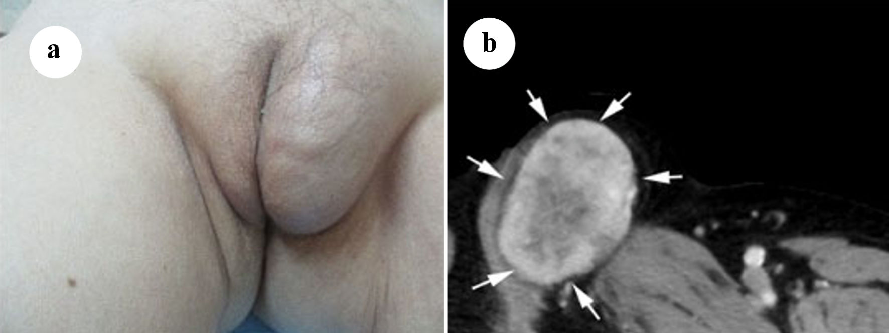
Figure 1. (a) Soft tissue mass on the left labia majora. (b) Axial computed tomography (CT) scan with intravenous contrast showing a mass in the left labia majora with intermediate density and enhancement (arrows).
Figure 2.
Figure 2. (a) Axial T1-weighted magnetic resonance imaging (MRI) of the tumor in the left labia majora revealed marginally higher signal intensity in the peripheral region and partially intermediate signal intensity in the central region than that in the surrounding muscle (arrows). (b) T2-weighted image revealed high signal intensity in the peripheral region and marked high signal intensity in the central region (arrows).
Figure 3.
Figure 3. Photomicrograph. The tumor is composed of spindle cells without nuclear atypia and fibrous stroma forming hemangiopericytomatous pattern (a) Hematoxylin and eosin stain (× 200). The tumor cells are focally positive for CD34 (b) and strongly positive for STAT6 (c) (× 200 immunohistochemical staining).